頰部 (Cheek)
頰部 (Cheek)
前區包含頰肌 (buccinator muscle)、頰脂肪墊 (buccal fat pad)、面神經 (facial nerve) 的頰支 (buccal branches),以及向下走行的腮腺管 (parotid duct)。
腮腺區 (parotid region) 以腮腺 (parotid gland) 與其下方深部的咬肌 (masseter muscle) 為主。
腮腺管 (parotid duct) 與面動脈 (facial artery) 是此區易於辨認的標誌。
下頜緣神經 (marginal mandibular nerve) 是下頜區的關鍵結構,因其一致地在頸闊肌 (platysma) 纖維的深面跨越面動脈 (facial artery)。
就解剖而言,頰部自耳的前緣延伸,內側受鼻、唇與頦所限,並自下頜 (mandible) 向上延伸至顴弓 (zygomatic arch) 與眶緣 (orbital rim)。然而,為了最佳地理解頰部的美容單位,最好將其描述為三個區域:前區 (anterior)、下頜區 (mandibular) 與咬肌—腮腺區 (masseter–parotid regions)。4,9,22
前區含有許多臉部表情肌。笑肌 (risorius) 非常表淺、大小不定,有時甚至缺如。顴大肌與顴小肌 (zygomaticus major and minor) 以及提上唇肌 (levator labii superioris) 與提上唇鼻翼肌 (levator labii superioris alaeque nasi) 起自顴骨 (zygomatic bone) 與眶緣 (orbital rim),並被眼輪匝肌 (orbicularis oculi muscle) 的纖維重疊。當翻起此區的皮膚時,顴小肌 (zygomaticus minor) 比其餘肌肉更表淺地呈現,其上覆僅有數毫米的皮下組織 (Figs. 1-6 and 1-23)。由於面神經 (facial nerve) 分支維持其走行與神經支配於肌肉的深面,只要剝離平面保持在肌肉平面的淺層,對這些分支的傷害便較不可能發生。
沿前頰部的內側面,面動脈 (facial artery) 可見於面靜脈 (facial vein) 的前方,它在跨越下頜後朝口角上行,並在該處發出唇支 (labial branches),隨後以角動脈 (angular artery) 形式橫越鼻唇溝 (nasolabial groove)。在鼻唇溝 (nasolabial groove) 內,藉由在皮下組織以及其上覆的顴大肌與顴小肌 (zygomaticus major and minor muscles) 肌性小束內剝離,可不太困難地觸及角動脈 (angular artery) (Fig. 1-19)。角動脈 (angular artery) 隨後在內眥韌帶 (medial canthal tendon) 正上方,藉由與頸內動脈 (internal carotid artery) 的眼動脈分支 (ophthalmic branch) 吻合,終止於內眥 (medial canthus) (Fig. 1-19)。此頰部區域最深層的結構為頰肌 (buccinator) 與提口角肌 (levator anguli oris muscles)。與其他臉部表情肌不同,頰肌 (buccinator) 是一塊相對厚實 (fleshy) 的肌肉。它位於一個下陷平面 (drop-down plane) 內,由大量脂肪組織覆蓋,後者常被稱為頰脂肪墊 (buccal fat pad) (Fig. 1-25)。頰肌 (buccinator) 被腮腺管 (parotid duct) 以及血管和上頜神經 (maxillary nerve) 的頰支所穿透,這些頰支前往供應頰黏膜 (buccal mucosa) 的感覺神經支配。4,9,22 頰脂肪墊 (buccal fat pad) 本身在技術上位於中頰部 (medial cheek)。此脂肪團輪廓分明,由一層薄的筋膜所包覆,在其表面可見面神經 (facial nerve) 分支跨越脂肪墊,仍位於 SMAS 的深面 (Fig. 1-6)。在上方,眶下神經 (infraorbital nerve) 分支成一束神經 (leash of nerves),以供應中頰部的感覺神經支配。眶上孔 (supraorbital foramen) 在大多數個體可輕易觸診到。
咬肌—腮腺區 (masseter–parotid region) 提供一個非常重要的解剖標誌——深部設置的咬肌 (masseter muscle)。咬肌為一塊由三叉神經 (trigeminal nerve) 支配的咀嚼肌,當咬緊牙關時可輕易視覺辨認與觸診。其強而有力的附著延伸於顴弓 (zygomatic arch) 與下頜支 (ramus of the mandible) 之間,而其前方的肌腱性緣 (anterior musculotendinous border) 為關鍵的臉部結構提供一個重要標誌。咬肌的後半部由腮腺 (parotid gland) 覆蓋。腮腺 (parotid gland) 包含於其自身的筋膜內,楔入於耳前下頜區 (preauricular mandibular region),並與咬肌筋膜 (masseteric fascia) 分隔——後者是頸深筋膜淺層 (superficial layer of deep cervical fascia) 的延續。在大多數個體中,咬肌的下前部可能被頸闊肌 (platysma) 的纖維重疊。
當在未經防腐處理的屍體標本上視察時,腮腺 (parotid gland) 呈現黃色,與活體病人非常相似。由於它相對接近皮膚本身,有時可能與皮下脂肪相混淆 (Fig. 1-23)。然而與皮下組織不同,腮腺 (parotid gland) 包含於一個有光澤、緊繃的筋膜鞘——腮腺筋膜 (parotid fascia)——之內,這可能有助於將其與脂肪區分。在腺體的前緣,可定位到腮腺管 (parotid duct),它先水平行走、然後向下進入頰脂肪 (buccal fat)。它一致地跨越咬肌 (masseter muscle),然後急轉朝向頰肌 (buccinator) 並穿透之,進入口腔,正對上頜第二臼齒 (upper second molar)。從其體表解剖而言,腮腺管 (parotid duct) 可定位於屏唇線 (tragolabial line) 22 與咬肌 (masseter muscle) 前緣的交會區域,此外還沿顴弓 (zygomatic arch) 下方約 2 cm 處。腮腺管 (parotid duct) 是一個顯著的結構,提供可靠的解剖標誌。由於腮腺管 (parotid duct) 位於 SMAS 的深面,它被面神經顴支 (zygomatic branch of the facial nerve) 的纖維所跨越,並被面神經上、下頰支 (upper and lower buccal branches of the facial nerve) 所夾擠 (Figs. 1-5, 1-6, and 1-23)。
面橫動脈 (transverse facial artery) 發自頸外動脈 (external carotid artery),與腮腺管 (parotid duct) 平行通過,介於腮腺管與顴弓 (zygomatic arch) 之間。它亦跨越咬肌 (masseter muscle) 的前緣。顳淺動脈 (superficial temporal artery) 在顴弓 (zygomatic arch) 下方、耳屏 (tragus) 正前方自腮腺筋膜 (parotid fascia) 穿出 (Fig. 1-27)。4,9,22 它通過腮腺 (parotid gland) 的後方與深面,是頸外動脈 (external carotid artery) 的終末分支之一。面動脈與面靜脈(靜脈位於動脈後方)在跨越下頜時亦行走於 SMAS 之下。面動脈與面靜脈可定位於咬肌 (masseter muscle) 的正前方。面動脈 (facial artery) 以迂曲 (tortuous) 的上行路徑行走,在年長者常更為明顯地迂曲。面神經 (facial nerve) 的下頜支 (mandibular branch) 在面動脈跨越下頜處上方約 5 to 10 mm 跨越面動脈。
下頜區 (mandibular region) 自咬肌 (masseter muscle) 的前緣延伸至頦。翻起此區皮膚後會遇到三塊主要肌肉。頸闊肌 (platysma) 位於淺筋膜 (superficial fascia) 內,並插入下唇的皮膚,與口輪匝肌 (orbicularis oris muscle) 的纖維融合。降口角肌 (depressor anguli oris) 起自下頜、插入口角,亦發出與頸闊肌 (platysma) 及口輪匝肌 (orbicularis oris) 的纖維融合的纖維。
面動脈與面神經的下頜緣支 (marginal branch of the facial nerve) 皆位於頸闊肌 (platysma) 纖維的深面。在大多數個體中,面神經的下頜緣支 (marginal branch) 維持在下頜緣 (mandibular rim) 上方的走行,跨越面動脈以供應唇與口的降肌群。雖然通常有兩條分支,但面神經的下頜支 (mandibular branch) 可能以單一神經存在,或具有多達四條分支 (Fig. 1-26)。
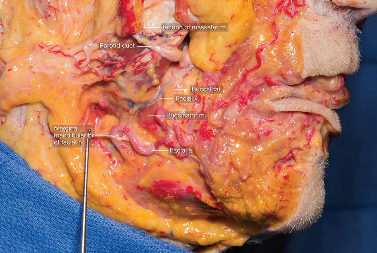
圖 1-5:面神經 (facial nerve) 的深部解剖,腮腺 (parotid gland) 上部已翻起 (Deep dissection of facial nerve with reflected upper portion of parotid gland)。
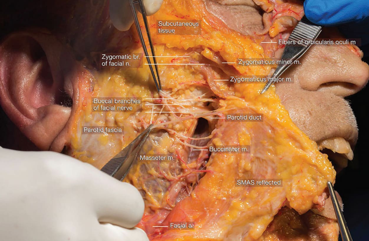
圖 1-6:SMAS 深面的面神經 (facial nerve) 解剖 (Dissection of facial nerve deep to SMAS)。
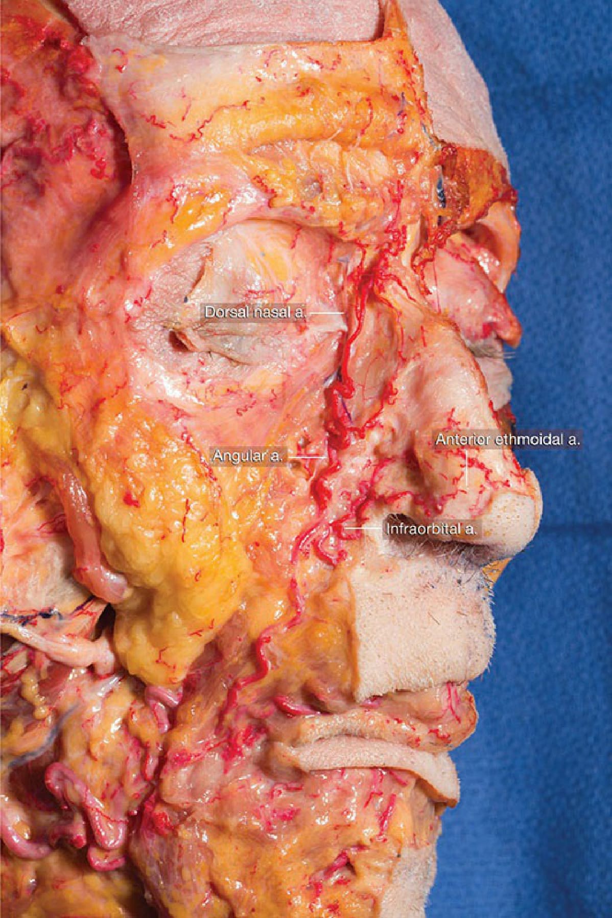
圖 1-19:解剖示範面動脈與角動脈 (facial and angular artery) 於鼻唇區 (nasolabial region) 內的上行 (Dissection demonstrating ascent of the facial and angular artery within the nasolabial region)。
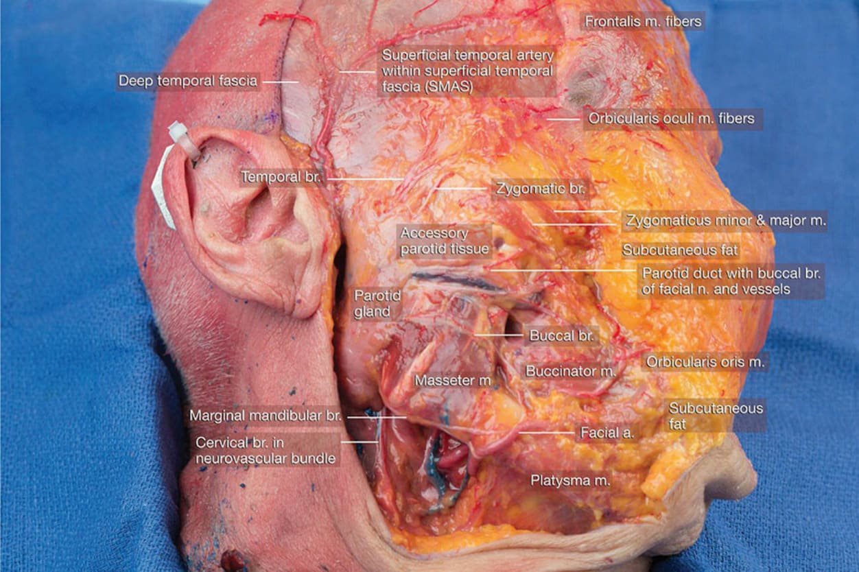
圖 1-23:外側臉部解剖,皮膚與皮下層向內側翻起 (Dissection of the lateral face with skin and subcutaneous layers reflected medially)。
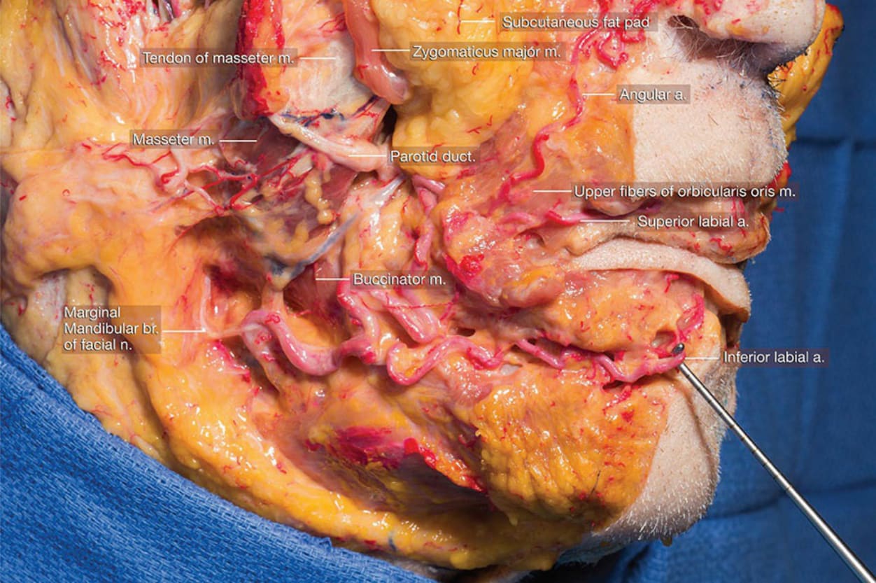
圖 1-25:中頰部與下頜區周圍區域解剖的外側觀 (Lateral view of dissection of regional anatomy around the mid cheek and mandibular region)。
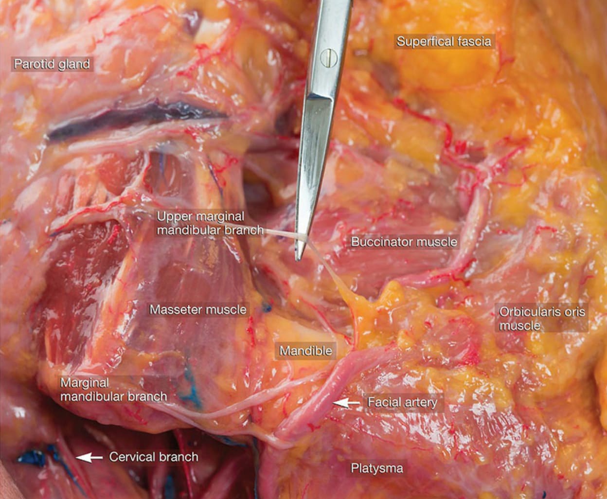
圖 1-26:解剖凸顯面神經下頜緣支 (marginal mandibular branch of the facial nerve) 及其與面動脈 (facial artery) 的關係 (Dissection highlighting the marginal mandibular branch of the facial nerve and its relationship to the facial artery)。
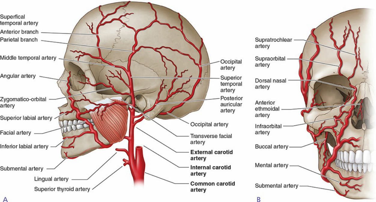
圖 1-27:圖示臉部的動脈型態與供應 (Diagram illustrating the arterial pattern and supply to the face)。