鼻 (Nose)
鼻 (Nose)
鼻分為鼻根 (root)、鼻背 (dorsum)、外側壁 (lateral walls)、鼻尖 (tip)、鼻翼 (alae) 與鼻小柱 (columella)。
大部分的鼻翼 (alae) 由皮膚與纖維脂肪組織 (fibrofatty tissue) 構成。
血液供應外側主要來自角動脈 (angular artery)、內側來自蝶腭動脈 (sphenopalatine artery),並有來自上唇動脈 (superior labial) 與眼動脈 (ophthalmic arteries) 的較小貢獻。
感覺神經支配源自上頜神經 (maxillary nerve) 的眶下分支 (infraorbital branch),以及前篩神經 (anterior ethmoidal nerve)(屬眼神經 ophthalmic nerve)的滑車下支 (infratrochlear) 與鼻外支 (external nasal branches)。
在鼻部進行手術的挑戰有兩方面。一方面,鼻在一個相當小的解剖界線之內,呈現由皮膚、軟骨 (cartilage) 與鼻黏膜 (nasal mucosa) 構成的複雜解剖。其次,鼻位於中臉部 (mid-face) 的位置,使美容結果格外受重視,這更加強調透徹理解解剖的重要性,以促進有效的手術修復與結果。在簡單的描述中,鼻可分為鼻根 (root)、鼻背 (dorsum,即鼻樑 bridge)、外側壁 (lateral side walls) 與鼻小葉 (lobule) (Fig. 1-17)。鼻小葉 (lobule) 進一步分為鼻尖 (nasal tip)、鼻尖下 (infra-tip) 與鼻翼 (alae)。由下方觀看時,鼻尖下小葉 (infra-tip lobule) 在前方呈現一個柔軟的三角形區域,一條向下延伸並分隔兩個鼻孔 (nostrils) 的鼻小柱 (columella)(鼻孔受鼻孔檻 nostril sills 所界定),以及外側的鼻翼基部 (alar base) 與鼻翼緣 (rim)。骨性錐體 (bony pyramid)、鼻中隔 (septum)、鼻翼軟骨 (alar cartilages) 與軟骨性穹窿 (cartilaginous vault) 共同構成鼻的主要結構支撐。
鼻骨 (nasal bones) 沿中線相互關節,並在外側與上頜骨的額突 (frontal processes of the maxillae) 關節。在上方,鼻骨與額骨的鼻突 (nasal processes of the frontal bone) 關節,在下方則與篩骨的垂直板 (perpendicular plate of the ethmoid bone) 關節。鼻骨 (nasal bones) 在上方最厚,但向下方則變薄,在該處易受損傷。外側軟骨 (lateral cartilages) 的上、下緣之間有重疊。骨性錐體 (bony pyramid) 上方的皮膚鬆弛、相當可動,並可輕易潛行剝離 (undermined)。22,23
外側軟骨 (lateral cartilages) 是鼻骨 (nasal bones) 的延續,其上方被鼻骨重疊,下方則被鼻翼軟骨外側腳 (lateral crura of the alar cartilages) 的上緣重疊。韌帶組織 (ligamentous tissue) 連接這兩處的懸垂部分。
鼻中隔 (nasal septum) 由骨、軟骨與軟組織構成,包含其所有相關節的顱顏骨性結構。鼻中隔軟骨(或稱四角形軟骨 quadrangular cartilage)錨定於篩骨的垂直板 (perpendicular plate of the ethmoid bone),並維持骨性中隔 (bony septum) 的結構完整性。膜性中隔 (membranous septum) 是一個軟組織複合體,由兩層被疏鬆結締組織 (loose connective tissue) 分隔的鼻前庭皮膚 (vestibular skin) 構成。降鼻中隔肌 (depressor septi muscle) 橫越膜性中隔 (membranous septum),並附著於鼻中隔軟骨 (septal cartilage) 的下緣。4,22
鼻小葉 (lobule) 是鼻最可動的部分,因其缺乏任何固定的軟骨關節。鼻小葉 (lobule) 的支撐來自由軟組織韌帶懸吊的成對鼻翼軟骨 (alar cartilages)。鼻翼 (ala) 的軟組織部分不含軟骨,而是藉由增厚的真皮 (dermis) 加以結構性維持,其下方並無皮下脂肪 (subcutaneous fat),使得在此區偵測理想的剝離平面具有挑戰性。
鼻周圍的關鍵肌肉包括皺眉肌 (procerus)、提上唇鼻翼肌 (levator labii superioris alaeque nasi)、鼻肌 (nasalis) 與降鼻中隔肌 (depressor septi muscles)。皺眉肌 (procerus) 從額肌 (frontalis muscle) 跨越鼻根 (root of the nose) 延伸,並與橫向走行的鼻肌 (nasalis muscle) 融合。重要的是須牢記鼻肌 (nasalis) 深面的平面與帽狀腱膜下平面 (subgaleal plane) 相連續,此平面可維持無血的剝離視野。提上唇鼻翼肌 (levator labii superioris alaeque nasi) 起自上頜骨 (maxilla),並發出纖維至上唇內側部與鼻翼外側。這些肌纖維最內側的部分稱為降鼻中隔肌 (depressor septi),它向下牽拉鼻中隔並維持氣道通暢 (Fig. 1-18)。
鼻接受豐富的血液供應,這是一項手術優勢,使皮瓣 (flap) 設計與方向得以靈活運用。雖然血液供應外側主要來自角動脈 (angular artery)、內側來自蝶腭動脈 (sphenopalatine artery),並有來自上唇動脈 (superior labial) 與眼動脈 (ophthalmic arteries) 的較小貢獻,但最大的血管貢獻源自頸外動脈系統 (external carotid system)。上、下唇動脈 (superior and inferior labial arteries) 是面動脈 (facial artery) 的分支,它們沿鼻唇溝 (nasolabial grooves) 內的外側面、以上行的角動脈 (ascending angular artery) 形式繼續前行,前往內眥吻合部位 (medial canthal anastomotic site)。角動脈 (angular artery) 發出許多小分支至外側壁、鼻翼與鼻背,並形成自由的與對側的吻合,最終藉由與鼻背動脈 (dorsal nasal artery) 的連結而終止 (Fig. 1-19)。此吻合點高度可預期,其一致的呈現使其成為皮瓣構建非常可行的血管蒂 (pedicle)。眉間 (glabella) 與前額中部由滑車上動脈 (supratrochlear artery) 供應,後者是眼動脈 (ophthalmic artery) 的分支,在鼻背與鼻尖的鼻部重建中亦是可靠的血管蒂 (Fig. 1-20)。在鼻骨深面,鼻外動脈 (external nasal artery) 浮現至鼻背 (Fig. 1-21)。它通常伴隨前篩神經 (anterior ethmoidal nerve) 的鼻外支,後者提供鼻背與鼻尖的感覺神經支配。眶下動脈 (infraorbital artery) 亦貢獻於此區周圍的血管吻合。靜脈回流遵循動脈供應的型態,並未顯示任何具重要意義的解剖。
鼻的感覺神經支配藉由三叉神經 (trigeminal nerve) 的眼支 (ophthalmic) 與上頜支 (maxillary divisions) 之分支達成。眼支供應鼻中線沿線的區域,而上頜支則經由眶下神經 (infraorbital nerve) (Fig. 1-20) 支配鼻翼、下外側壁與鼻小柱。鼻根 (root) 與上鼻樑 (upper nasal bridge) 連同上外側壁,則由滑車下神經 (infratrochlear nerve) 供應,後者自內眥韌帶 (medial canthal tendon) 上方以朝內側的方向接近鼻。
耳 (Ear)
外耳分為耳廓 (auricle,即耳殼 pinna)、外耳道口與外耳道 (external auditory meatus and canal),以及位置較深的鼓膜 (tympanic membrane) 之外表面。
耳的血液供應源自顳淺動脈 (superficial temporal artery) 的上、下耳支 (superior and inferior auricular branches),以及上頜動脈 (maxillary artery) 的耳深支 (deep auricular branch)。
外耳接受來自重疊的腦神經與頸神經 (cranial and cervical nerves) 的豐富感覺神經支配。
耳顳神經 (auriculotemporal nerve) 行走於顳淺血管 (superficial temporal vessels) 的後方,並供應耳廓 (auricle) 的前部與前耳輪 (anterior helix)。
耳顳神經 (auriculotemporal nerve) 位於顳淺動脈與靜脈 (superficial temporal artery and vein) 的後方,並在橫越腮腺 (parotid gland) 時自腮腺上方筋膜 (superior parotid fascia) 穿出。
乳突區 (mastoid area) 由經由枕小神經 (lesser occipital nerve) 而來的 C2、C3 腹側支 (ventral rami) 供應。耳甲 (concha) 由腦神經 VII、IX 與 X 的不定且重疊的神經支配所供應,這些神經亦供應外耳道後部、鼓膜以及耳後溝 (posterior auricular sulcus)。
對皮膚外科醫師而言,理解耳的結構對於修復大型與小型缺損皆屬必要。在進行潛行剝離 (undermining)、執行直接縫合 (primary closure) 或進行組織移動 (mobilization) 時,瞭解皮膚厚度、彈性、與其下軟骨之關係,以及灌注型態的變異,有助於產生最有效的修復。
外耳分為耳廓 (auricle,即耳殼 pinna)、外耳道口與外耳道 (external auditory meatus and canal),以及位置較深的鼓膜 (tympanic membrane) 之外表面。22 耳廓 (auricle) 由一個複雜的軟骨支架構成,並摺疊成各種皺褶與溝槽。軟骨由緊密貼附、皮下組織極少的皮膚覆蓋,常常完全沒有真皮下脂肪 (subdermal fat)。皮膚在前方緊繃,後方則提供稍多一些的彈性。耳廓最下方的部分為耳垂 (lobule),沒有軟骨基底,由皮下脂肪與皮膚構成。耳垂上方延伸出兩道明顯的彎曲:(1) 外側的耳輪 (helix)——一道向前彎曲的皺褶,自耳垂向後上延續至耳屏 (tragus) 上界附近,並在該處與耳輪腳 (crus of the helix) 融合;以及 (2) 對耳輪 (antihelix),藉由一道稱為舟狀窩 (scaphoid fossa) 的溝槽與耳輪分隔。耳屏 (tragus) 是耳廓軟骨的前向延伸,藉由屏間切跡 (intertragal space) 與對耳屏 (antitragus) 分隔。一道稱為耳甲 (concha) 的深凹溝通向外耳道口 (external auditory meatus)。耳甲 (concha) 進一步細分為較上方的凹陷——耳甲艇 (cymba),以及較下方、較大的凹陷——耳甲腔 (cavum) (Fig. 1-22)。4,9,22
雖然存在變異,但在其標準解剖位置上,耳位於外側,大致介於眉毛與鼻基底之間,耳輪 (helix) 突出於對耳輪 (antihelix) 之外。韌帶纖維將耳廓連接至顱骨,並含有退化的內在肌 (rudimentary intrinsic muscles)。外在肌 (extrinsic muscles) 臨床意義不大,但有助於注意的是,這些臉部表情肌——耳前肌、耳後肌與耳上肌 (anterior, posterior, and superior auricular muscles)——皆包含於 SMAS 之內,並由面神經 (facial nerve) 的分支支配。
外耳道口與外耳道 (external auditory meatus and canal) 的長度為 2.5 to 3.5 cm。耳道本身兼具骨性與軟骨性部分。22 在外側,軟骨性成分與耳廓軟骨 (auricular cartilage) 相連續,而在內側則附著於骨性外耳道口 (bony meatus)。軟骨性部分大多存在於耳道的下方。在上方,耳道由顳骨鱗部 (squamous temporal bone) 所界定。耳道真正的骨性部分穿行於顳骨的鱗部與鼓部 (squamous and tympanic parts) 之間。在外耳道口的外側部周圍,皮膚較厚,具有皮脂腺、耵聹腺 (cerumeniferous glands) 與毛髮。骨性部分含有非常薄的一層上皮,且無毛髮與腺體。具特別臨床意義的是耳道軟骨性部分內的裂隙。這些隨機排列的裂隙稱為 Santorini 裂 (fissures of Santorini),為發展中的皮膚癌 (skin cancers) 向周圍組織擴散提供潛在的途徑。
耳豐富的血液供應源自顳淺動脈 (superficial temporal artery) 的上、下耳支 (superior and inferior auricular branches),以及上頜動脈 (maxillary artery) 的耳深支 (deep auricular branch)。此外,耳後動脈 (posterior auricular artery)——頸外動脈 (external carotid artery) 的一條分支——供應耳的後部。由於皮下脂肪稀少,動脈分支在皮膚內排列為單層血管。靜脈型態與動脈供應相對應,回流經由顳淺靜脈 (superficial temporal) 與下頜後靜脈 (retromandibular veins) 進行。
外耳接受來自重疊的腦神經與頸神經 (cranial and cervical nerves) 的豐富感覺神經支配。三叉神經 (trigeminal nerve) 的下頜支 (mandibular division) 發出耳顳神經 (auriculotemporal nerve),後者行走於顳淺血管 (superficial temporal vessels) 的後方,並供應耳廓 (auricle) 的前部與前耳輪 (anterior helix)。此外,耳顳神經 (auriculotemporal nerve) 供應耳道的前壁與上壁,以及鼓膜 (tympanic membrane) 外表面的一部分 (Fig. 1-13)。藉由牢記耳顳神經 (auriculotemporal nerve) 位於顳淺動脈與靜脈 (superficial temporal artery and vein) 的後方、且其在下方橫越腮腺 (parotid gland) 時自腮腺上方筋膜 (superior parotid fascia) 穿出,可限制對其的傷害。耳大神經 (great auricular nerve)(C2、C3 腹側支)供應耳廓內側面的大部分以及耳廓外側面的後部。這將包括耳輪 (helix) 與對耳輪 (antihelix) 的大部分。乳突區 (mastoid area) 亦由 C2、C3 腹側支供應,但其神經支配是經由枕小神經 (lesser occipital nerve) 而來。耳甲 (concha) 由腦神經 VII 不定地支配,外耳道口則由腦神經 IX 與 X 支配。4,9,22 這些腦神經亦供應外耳道後部、鼓膜以及耳後溝 (posterior auricular sulcus)。
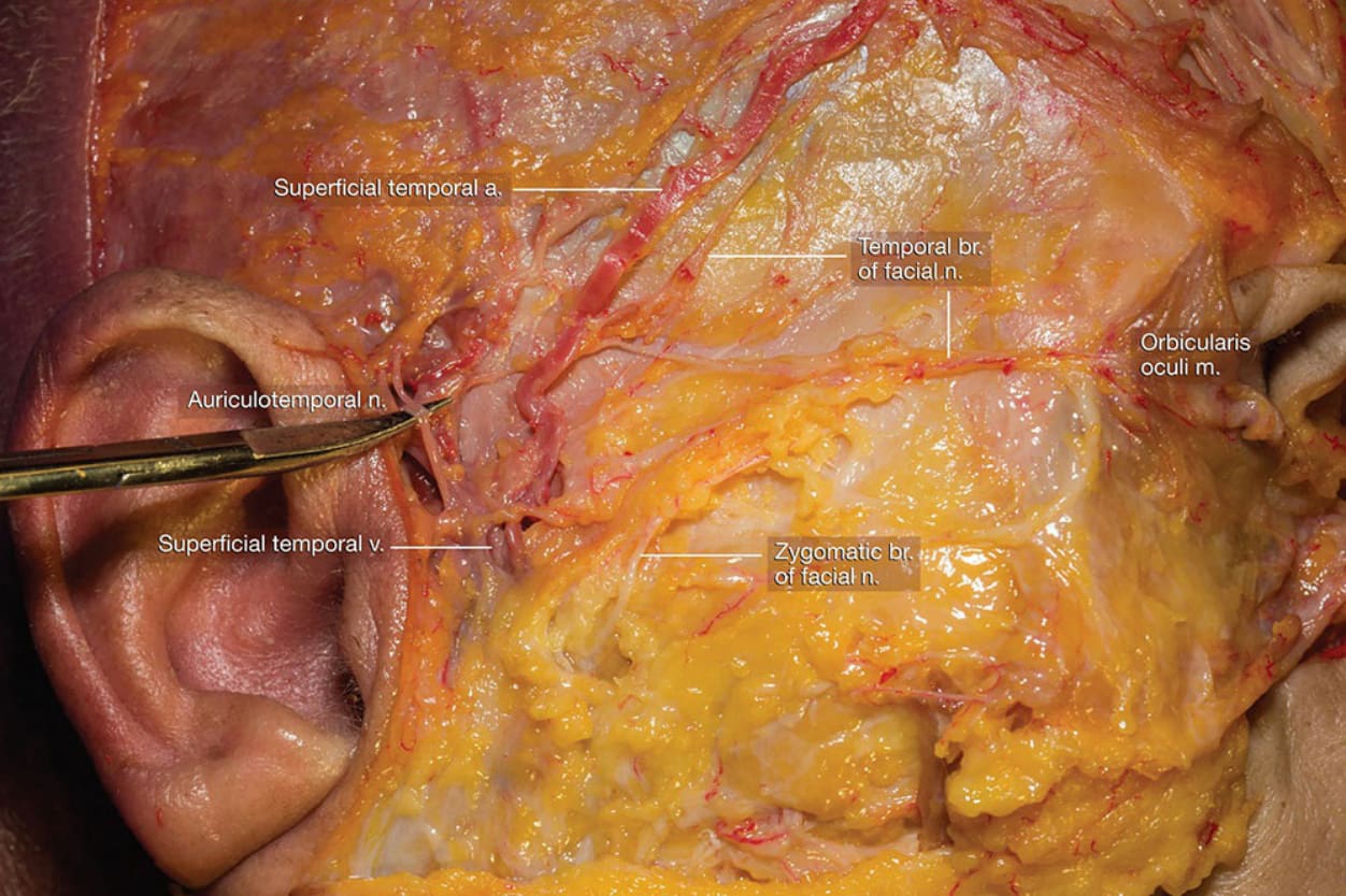
圖 1-13:顳區 (temporal region) 解剖,凸顯耳顳神經 (auriculotemporal nerve) (Dissection of temporal region highlighting the auriculotemporal nerve)。
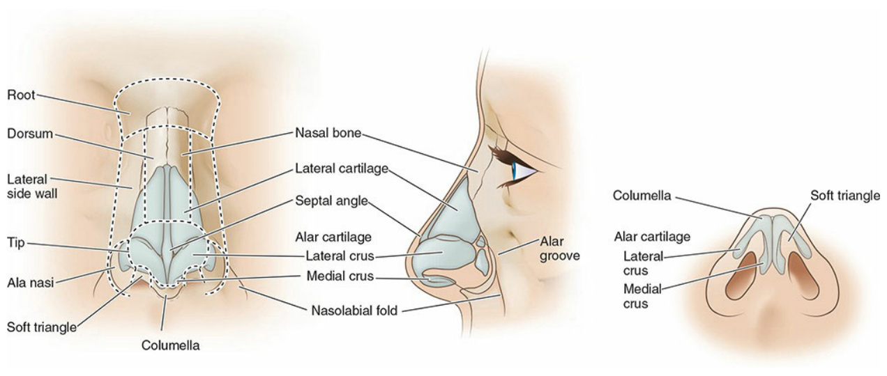
圖 1-17:圖示鼻的基本解剖 (Diagram illustrating basic anatomy of the nose)。

圖 1-18:圖示鼻周圍的深部解剖 (Diagram illustrating deeper anatomy around the nose)。
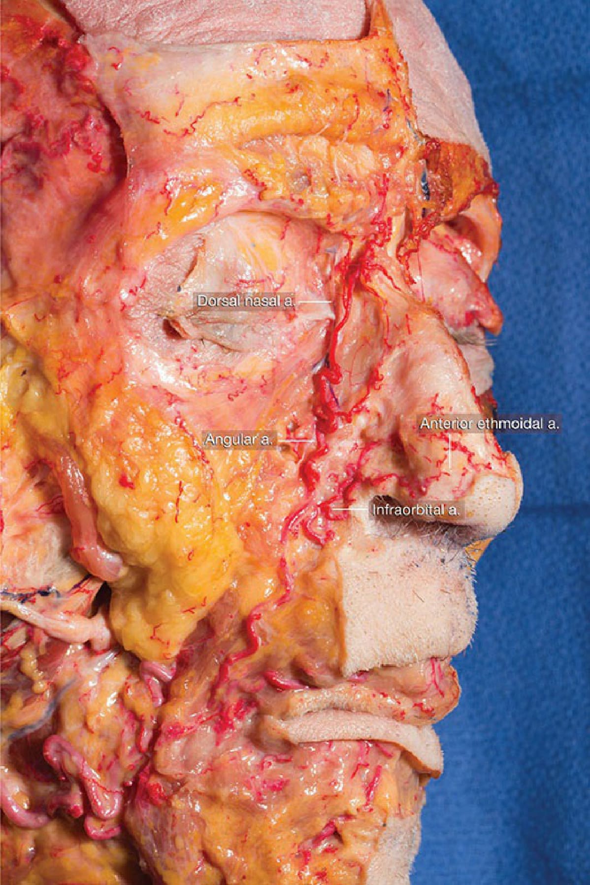
圖 1-19:解剖示範面動脈與角動脈 (facial and angular artery) 於鼻唇區 (nasolabial region) 內的上行 (Dissection demonstrating ascent of the facial and angular artery within the nasolabial region)。
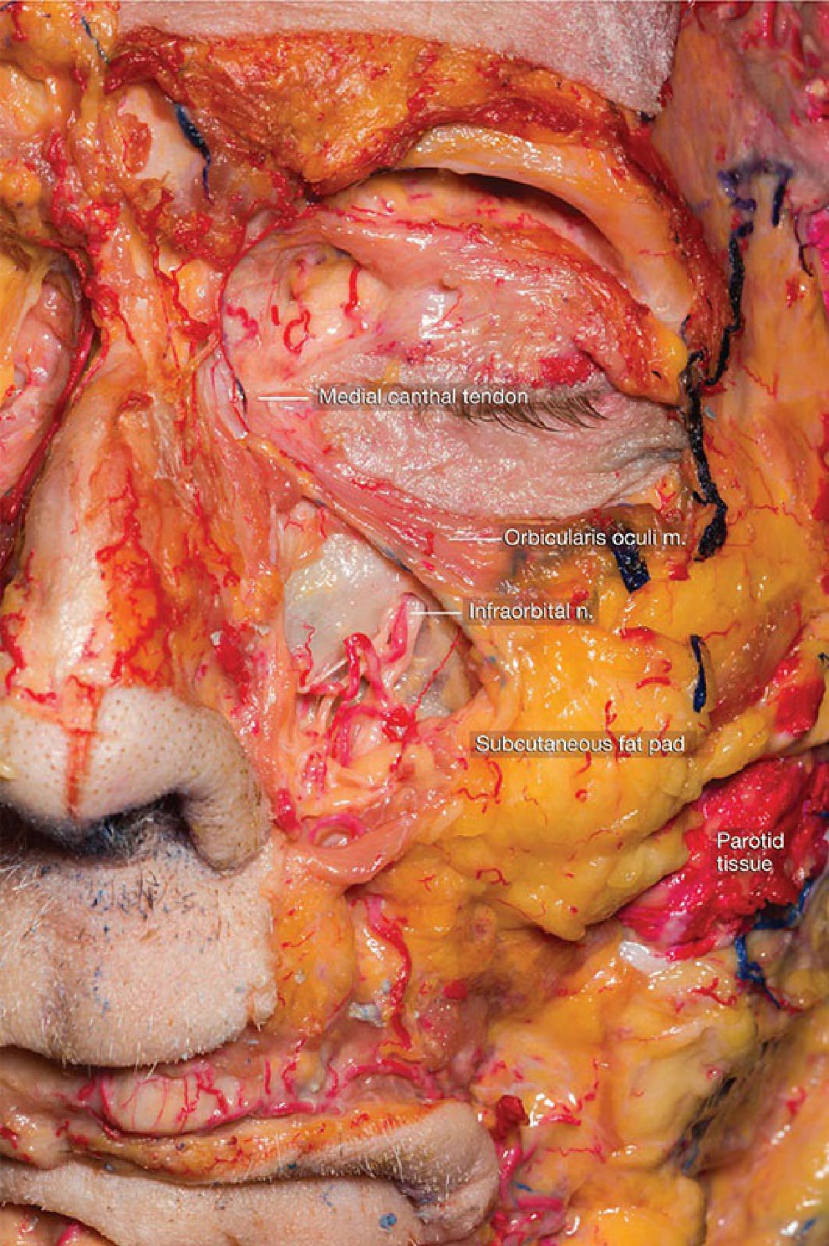
圖 1-20:左前頰部解剖,凸顯眶下神經 (infraorbital nerve) (Dissection of the anterior left cheek highlighting the infraorbital nerve)。
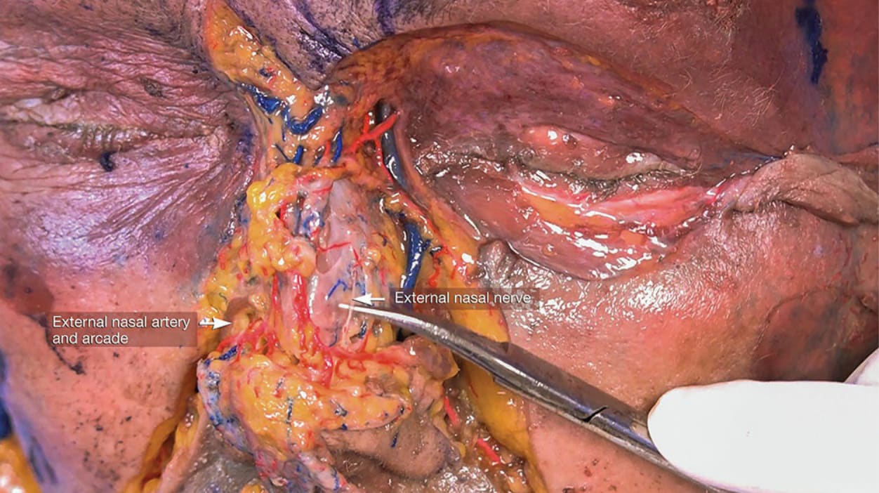
圖 1-21:前鼻部淺層解剖,示範鼻外神經與血管 (external nasal nerve and vessels) (Superficial dissection of anterior nose demonstrating the external nasal nerve and vessels)。
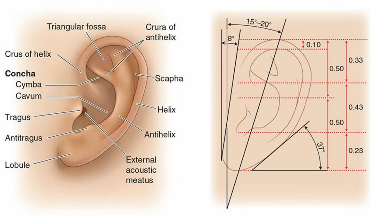
圖 1-22:圖示外耳的基本解剖 (Diagram illustrating basic anatomy of the external ear)。