Introduction
CHAPTER 77 Approaches to Erythema
and Telangiectasias
Margaret A. Weiss Anne M. Mahoney
SUMMARY
Erythema and telangiectasias are a common patient complaint.
The mainstay of therapy is laser and IPL treatments; IPL confers the advantage of
larger spot sizes, and thus more efficient treatments.
Patients should always understand that multiple treatments will be needed, and
maintenance therapy is generally required.
Beginner Tips
Regardless of the device chosen, a slight amount of overlap of pulses is necessary to
achieve uniform improvement of the treated area.
KTP causes more swelling and bruising compared to PDL and IPL.
PDL is safer than KTP in those with darker skin types given increased depth of
penetration.
Do not use IPL on those with tanned or sunburned skin.
There should be approximately 10% overlap with IPL to avoid a “stamping”
appearance following treatment.
Expert Tips
Telangiectasias of the nose can be very treatment resistant; recurrence rates are quite
high, necessitating maintenance treatments.
In many patients, it can be difficult to permanently eliminate alar telangiectasias
altogether; caution patients from the outset that maintenance treatments are often necessary.
Another factor for the physician to consider when treating large areas is the wear and
tear to the laser itself—larger areas consume more of the dye kit and cryogen.
Don’t Forget!
The neck and chest skin have a thinner epidermis and dermis compared to the face, so
typically gentler settings should be used.
It is important to become familiar with the device you choose to use. Devices of the
same wavelength do not necessarily have interchangeable settings, and the time to development of erythema or purpura following a single pulse varies between devices. Also, service of the laser (such as replacement of a dye kit) may influence the performance and actual energy delivered.
Pitfalls and Cautions
Do not pulse stack with a 1,064-nm laser, which increases the risk for scarring.
1,064 nm has the greatest risk for causing retinal damage because of its deep
penetration, so caution should be exercised near the eye with appropriate shielding.
Many laser experts avoid 1,064-nm laser treatments on the nasal ala due to an
increased risk for depressed scarring.
CHAPTER 77 Approaches to Erythema
and Telangiectasias
INTRODUCTION
Prominent erythema and telangiectasia of the face, neck, and chest are frequent complaints and reasons for presentation to the dermatology office. The most common etiologies of these signs are rosacea and photodamage, with less common reasons being alcoholism, medication use, connective tissue diseases, and genetic disorders, such as hereditary hemorrhagic telangiectasia. Sun exposure damages and weakens collagen and elastin, and cumulative exposure results in vascular ectasias and physical signs of erythema and telangiectasias.
UV photodamage is believed to contribute to the development of rosacea. Rosacea symptoms typically include frequent flushing associated with facial erythema, telangiectasias, papules, and pustules. Patients with CREST (calcinosis, Raynaud’s phenomenon, esophageal dysmotility, sclerodactyly, and telangiectasia) often present with what are classically referred to as mat-like telangiectasias. These are most prominent on the face, neck, and hands (Figs. 77-1 and 77-2).
IPL at 30 J/cm2 with pulse width of 2.4 ms, 15-ms delay, and 5 ms with significant improvement. The patient receives periodic laser treatment to maintain clearance of the mat telangiectasias, and was diagnosed with CREST syndrome at age 40.
Connective tissue disease and other systemic illnesses may cause facial erythema and telangiectasias; patients with polycythemia vera have been referred in the past for
laser treatments (Figs. 77-3 and 77-4). Medications that can cause erythema include niacin (rather than nicotinamide), calcium channel blockers, cyclosporine, rifampin, vasodilators such as nitroglycerin, and others. It is also important to keep in mind that topical and intralesional steroids can cause telangiectasias and erythema. Intrinsic aging of the skin causes collagen breakdown and visible telangiectasias. Lastly, repeated trauma to the face will also induce localized erythema, and ultimately vascular dilation. Telangiectasias frequently develop in and around surgical scars, particularly those that are under tension.
with 10-mm spot, 8.6 J/cm2, and 10 ms. The pulse count ranged from 153 to 160 pulses per session.
Currently, the most effective treatment of erythema and telangiectasias is by lasers and intense pulsed light (IPL); however, other therapies play a role as well. Patients with erythema and telangiectasias should be instructed on a gentle skin care routine of using lukewarm water, mild cleansers, and moisturizers. Prescription topical agents for erythematotelangiectatic rosacea include metronidazole gel, lotion and cream, azelaic acid foam and gel, and ivermectin cream. Although the former agents are more effective for papulopustular rosacea than erythema and flushing, topical agents may be included as adjunctive anti-inflammatory approaches for rosacea patients.
Brimonidine gel (Mirvaso, Galderma, Lausanne, Switzerland) is an α-2 adrenergic agonist which causes vasoconstriction and can be used for erythema of varying etiologies. When this agent was initially on the market there was much enthusiasm for its potential efficacy, though in clinical practice marked rebound erythema has occurred with cessation of use, even after 1 to 2 days of application; such rebound erythema may extend to areas beyond the area of direct application. Because of these side effects, the popularity of brimonidine has declined. Results of studies on topical oxymetazoline are currently under review by the Food and Drug Administration.
Cosmeceuticals can be used to reduce erythema and telangiectasias.1 Topical and oral nicotinamide, green tea, feverfew, and licorice root all have been shown to be possibly beneficial for erythema via varying anti-inflammatory mechanisms. Green tea
has also been shown to possibly protect against ultraviolet B (UVB) damage.
Historically, electrocautery was used to treat telangiectasias. This has fallen out of favor given the damage to the epidermis when trying to reach underlying vessels, and the risk of resultant scarring. In addition, while the immediate response to cautery appears successful, recurrence is common.
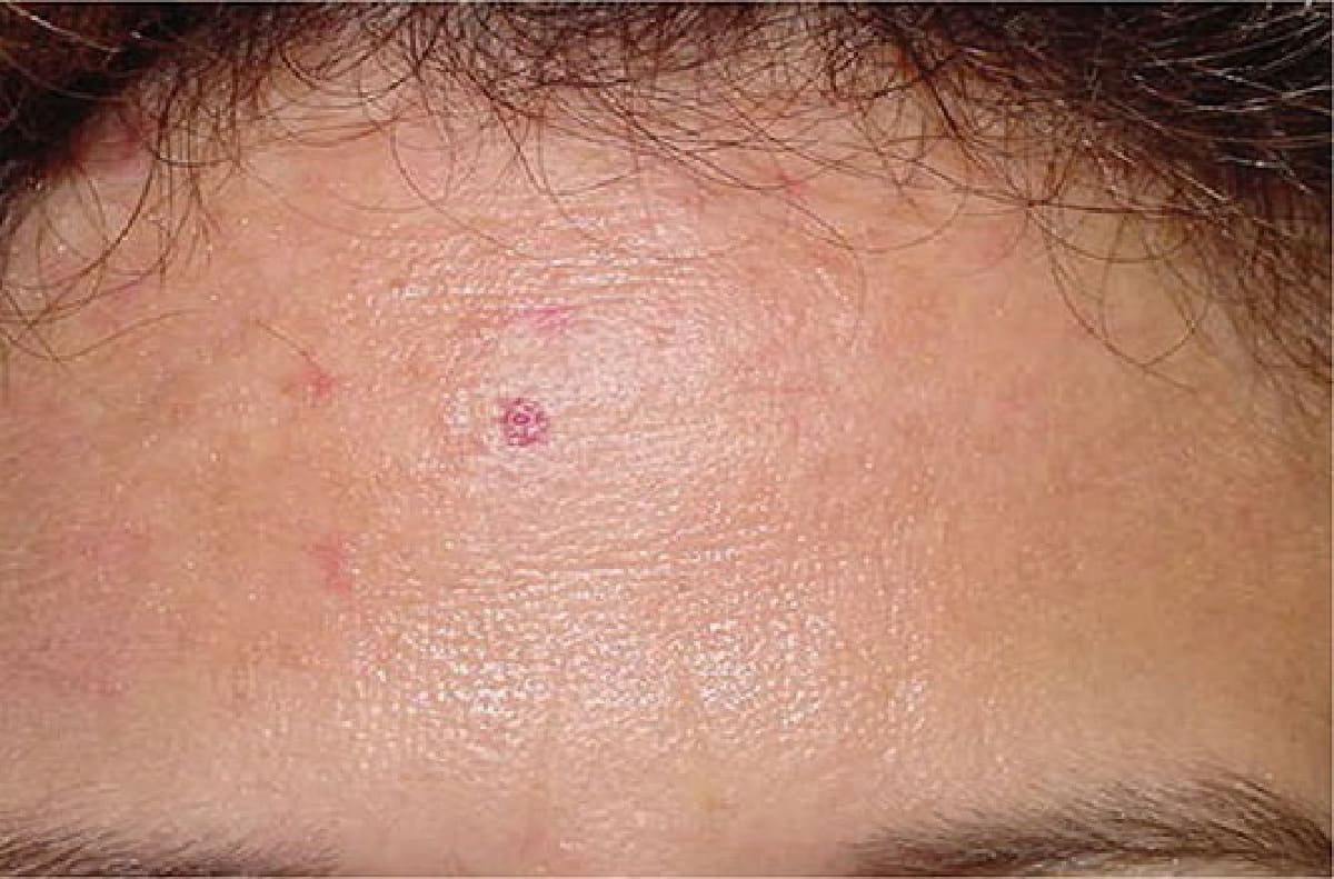
Figure 77-1. A 30-year-old female with a history of Raynaud’s presented for treatment of telangiectasias. She was noted to have mat telangiectasias on the forehead.
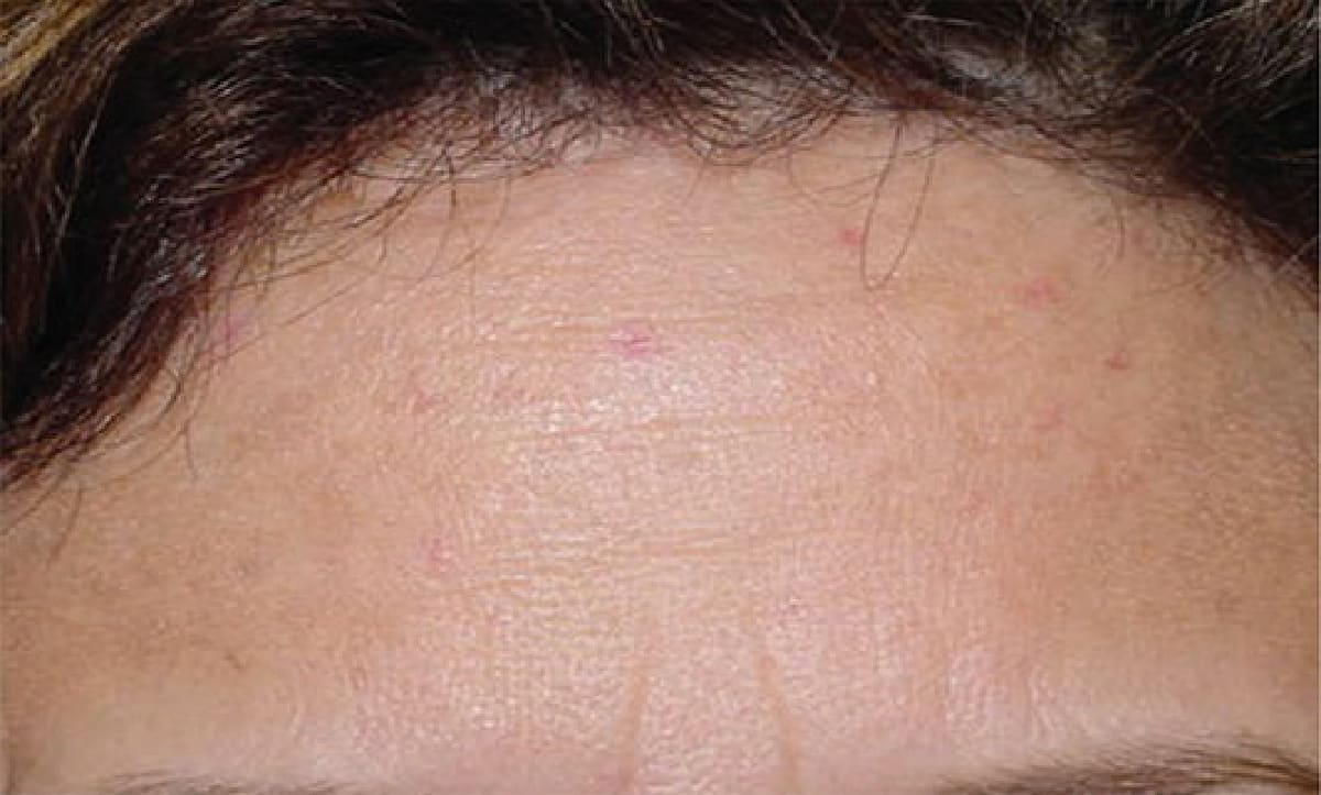
Figure 77-2. The same patient was treated with one session of 1,064 nm, 90 J/cm2, and 7 ms followed by the 590-nm
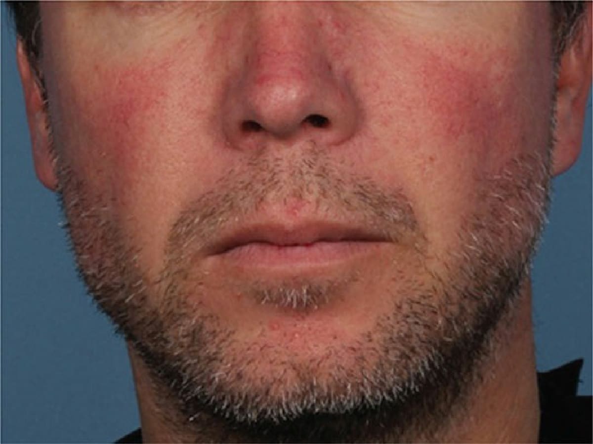
Figure 77-3. A 40-year-old male presented for treatment of redness of the face. Several years earlier, the patient had presented for the same complaint, and was noted to be diffusely erythematous and diaphoretic. He was referred to his primary care physician for workup of systemic illness, and was found to have polycythemia vera. The patient was started on aspirin 81 mg and phlebotomy. On reevaluation, the patient was much less red, but demonstrated prominent telangiectasias on the cheeks, nose, and chin and diffuse facial erythema.
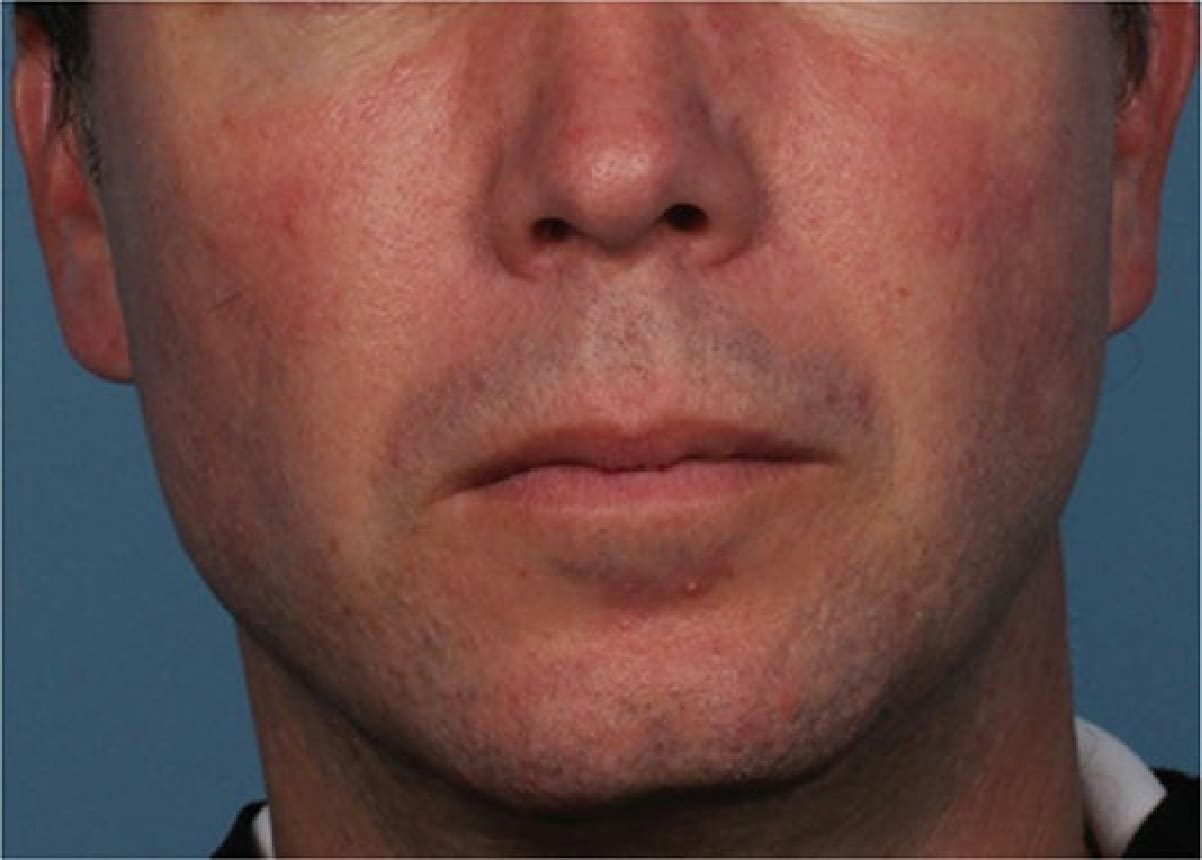
Figure 77-4. Marked improvement in erythema after receiving three treatments using the Cutera Excel V (535 nm)