Split-thickness skin grafting
Split-thickness skin grafting
First performed for the treatment of vitiligo by Haxthausen in 1947, STSG is considered one of the most effective methods of inducing repigmentation.26 This technique was refined further in 1964 by Behl, who was the first to describe the use of thin Thiersch grafts.27 Later in 1996, Kahn and Cohen utilized a motorized dermatome to obtain ultrathin STSGs for vitiligo surgery.14
Using this approach, a thin to ultra-thin STSG is harvested from the DS and temporarily placed in a sterile Petri dish filled with NS while DS dressings are placed and the RS is prepared by either laser or dermabrasion. The graft is then removed from the NS, placed dermal side down over the RS, and covered with appropriate dressings.26 A slide may be placed underneath the graft while it is being cut to more easily differentiate between the epidermal and dermal sides (Fig. 52-4). Alternatively, a marking pen may be used. The dermal side can also be distinguished by visualization of fibrin clots as well as inward curling. On digital microscopy, creasing is seen on the epidermal side.
The STSG technique is a desirable option when treating large areas, challenging areas such as the eyelids, areola, and genitals, and leukotrichia. This method is effective, and pigment return is fast and uniform. STSG is an in-office procedure, not requiring reagents, a lab, or expensive equipment. Drawbacks include hyperpigmentation, especially peripheral halo pigmentation, although this can be minimized by obtaining a DS graft 10% to 20% larger than the RS site to account for
graft contracture during healing. Milia and inclusion cysts may occur 2 to 4 months following the procedure,26 as well as peripheral beading, the inward curling of the graft rim during healing. Composite film and graft units, consisting of a semipermeable film extending past the graft edges, may be utilized to reduce the risk peripheral beading.28 In addition to graft hypertrophy, pincushioning, manifesting as a tire patch or stuck on appearance, can be seen when thicker grafts are placed. Graft rejection and DS scarring may also occur.26
Mesh grafting Mesh grafting is a modification of STSG that can be implemented to amplify graft size. Small slits are made throughout the graft to achieve a mesh-like appearance, thereby increasing graft size while also providing a route for exudative drainage at the RS.26 DS scarring may occur, and peripheral beading can be seen with thicker grafts.26
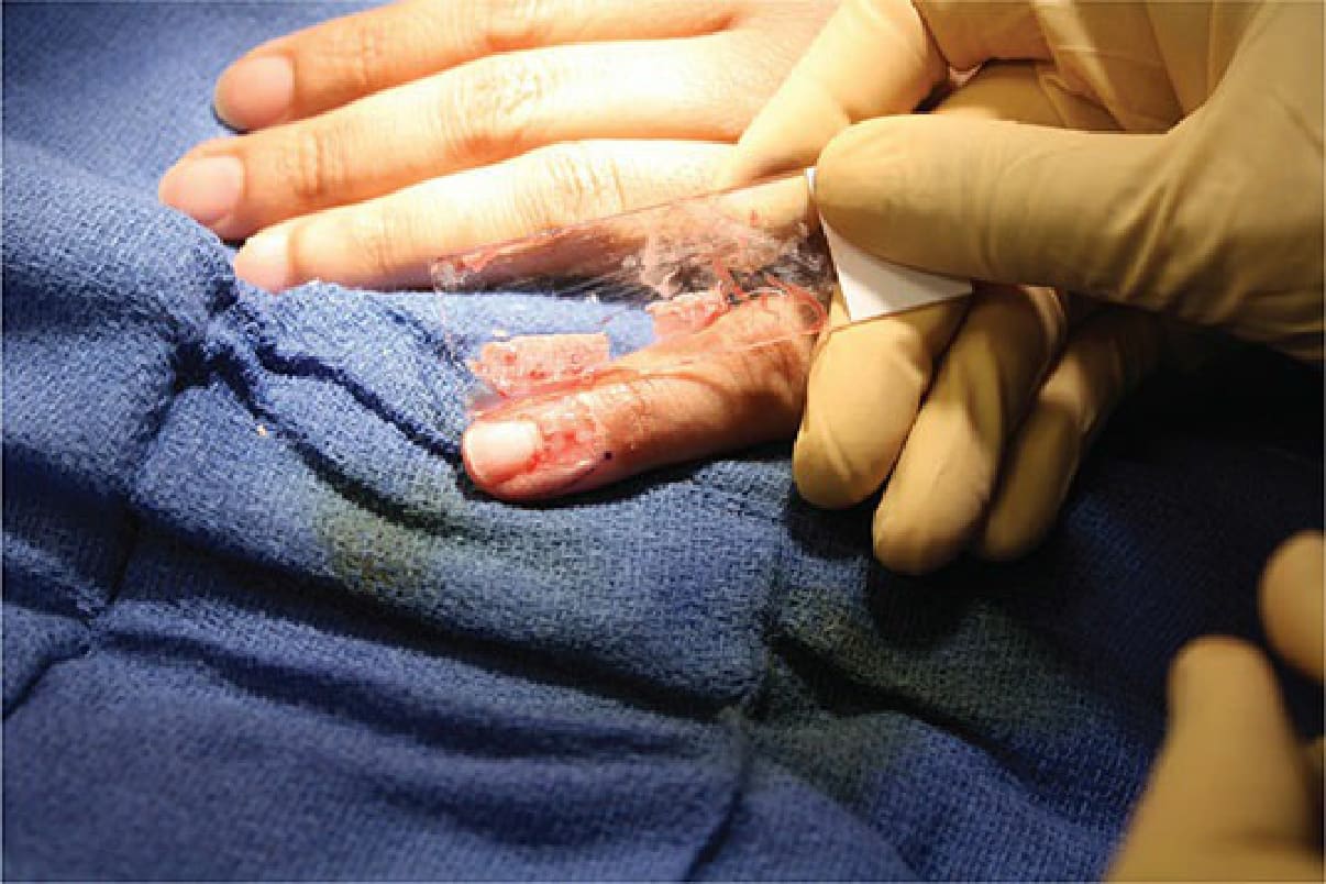
Figure 52-4. Placement of an STSG on the distal fingertip in a research patient using a glass slide.