Donor site preparation
Donor site preparation
Various techniques are employed to harvest tissue from the DS for cellular and tissue grafting. The main methods involve thin to ultra-thin skin grafts, suction blisters, and punch biopsies. Using a shaving technique, thin (0.125–0.250 mm)14 to ultra-thin (less than 0.125 mm)15 skin grafts can be harvested using either a sterile razor blade held by hemostats, a Weck blade, a Silver’s grafting knife, or a motorized dermatome.13,14,16 Maximal operator control of graft size and thickness is obtained with a sterile razor blade, but this method requires a significant degree of skill. The Weck blade allows for uniform graft thickness, but does not allow for adjustment of graft depth. In contrast, the Silver’s grafting knife allows for adjustment of depth. Motorized dermatomes provide uniform grafts and require very little user skill, but are more expensive.17 Appropriate grafts are transparent and float when placed in sterile saline.18 Curling of the graft edges indicates that the specimen is thick. This can be solved by either reharvesting the graft or by using higher concentrations of trypsin and longer incubation times if cell separation is being performed.
Punch biopsies, typically used for minigrafting or test spots, can also be used to harvest tissue. Equal-sized punch biopsies, usually 1 to 1.5 mm in diameter, are used to take tissue from the DS. Suction blisters are another method of obtaining tissue from the DS. This involves using a Luer lock disposable syringe with a three-way stopcock and pulling the plunger to create a pressure between 30 and 40 mmHg over a period ranging from 15 minutes to 3 hours (Fig. 52-1). This creates a blister, with separation of the skin at the DEJ (Fig. 52-2). Youthful skin requires higher suctioning pressures, whereas more mature skin requires lower suctioning pressures secondary to increased fragility due to decreased elastic fibers in the dermis.19 Sterile scissors are used to remove the blister, which is either transferred directly or manipulated and then transferred to the RS.11 The speed of blister formation can be accelerated by injection of sterile normal saline (NS)
or using heat.19 This method is not associated with scarring, as separation occurs at the DEJ. However, if excessive suction is employed, hemorrhagic blisters may develop, which may be too thick for use.
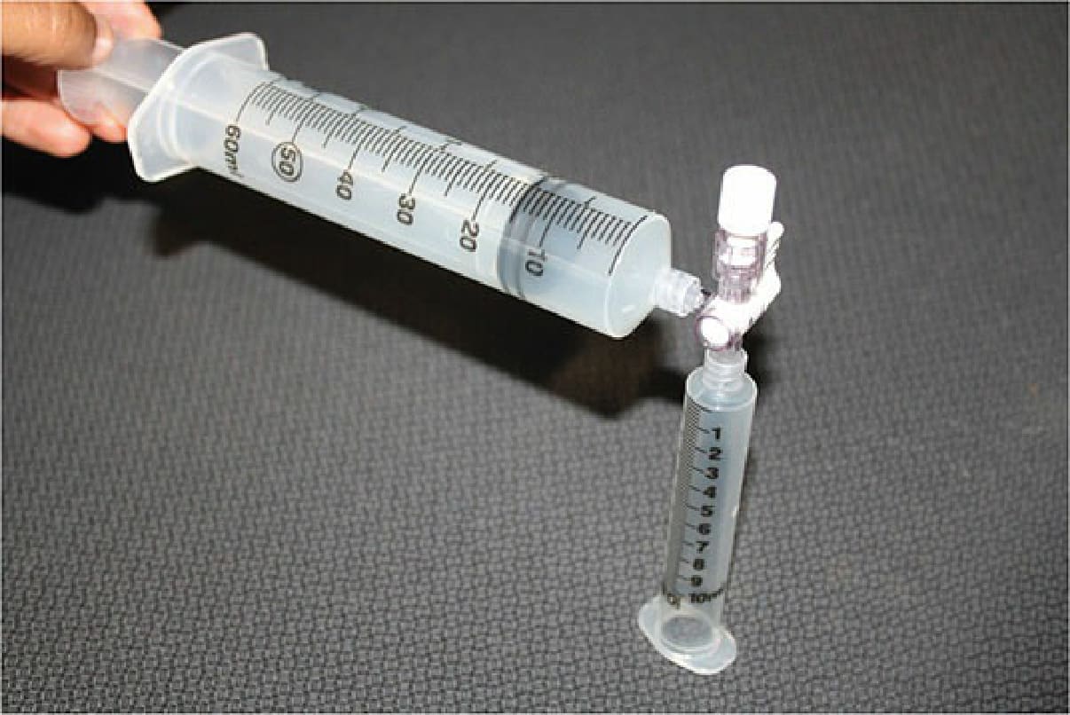
Figure 52-1. Suction blister apparatus using a Luer lock disposable syringe with a three-way stopcock and pulling plunger.
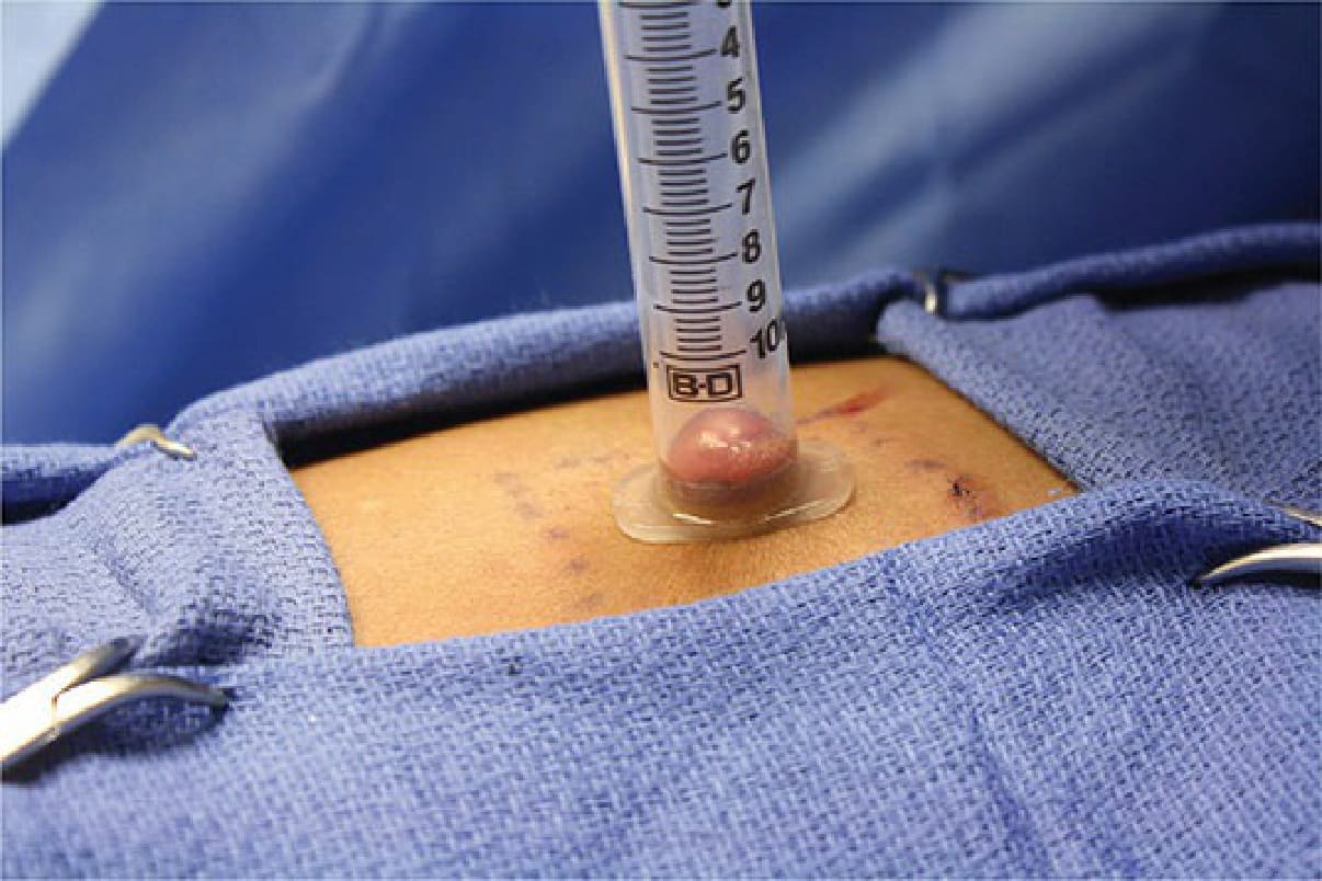
Figure 52-2. Suction blister induction.