CLINICAL PRESENTATION
CLINICAL PRESENTATION
Epidermal inclusion cysts present as soft subcutaneous nodules, sometimes with a visible punctum (or puncta), and an associated yellow–white hue (Figs. 50-1 and 50-2). These contain macerated keratin, and cyst contents are most frequently white or yellow, though they can be brown or gray-colored as well. Cyst size and location are variable, but they are most frequently found on the face and trunk, and are typically a few centimeters in size, depending on location. Primary milia are usually 1- to 3-mm white papules, most commonly occurring around the eyelids and central face (Fig. 50-3).
Clinically, pilar cysts are indistinguishable from epidermal cysts, though they occur primarily on the scalp and may be more firm to palpation. This increased firmness corresponds to the thick wall of the pilar cyst, which also makes them more resistant to rupture. Unlike epidermal cysts, a punctum is usually not seen with trichilemmal cysts (Fig. 50-4).8
Compared to epidermal cysts, steatocystomas tend to be smaller, softer acneiform lesions (usually less than 1 cm) occurring on the chest and axillae (Fig. 50-5).2 Dermoid cysts appear similar to epidermal cysts, but typically occur in infants and are located along embryonic fusion planes.3,9 Inflamed variants of each of these subtypes can occur, and will appear erythematous, warm, and tender due to the inflammatory reaction to the ruptured cyst wall and contents. Other common mimickers of epidermal cysts include lipomas, juvenile xanthogranulomas, and reactive lymph nodes.

Figure 50-1. Epidermal inclusion cysts present as soft subcutaneous nodules, sometimes with a visible punctum (or puncta), and an associated yellow–white hue.
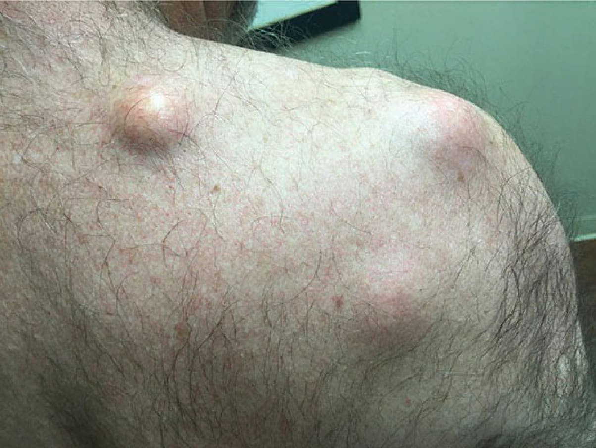
Figure 50-2. Epidermal inclusion cysts may become fairly large, and are in variable proximity to the skin surface.
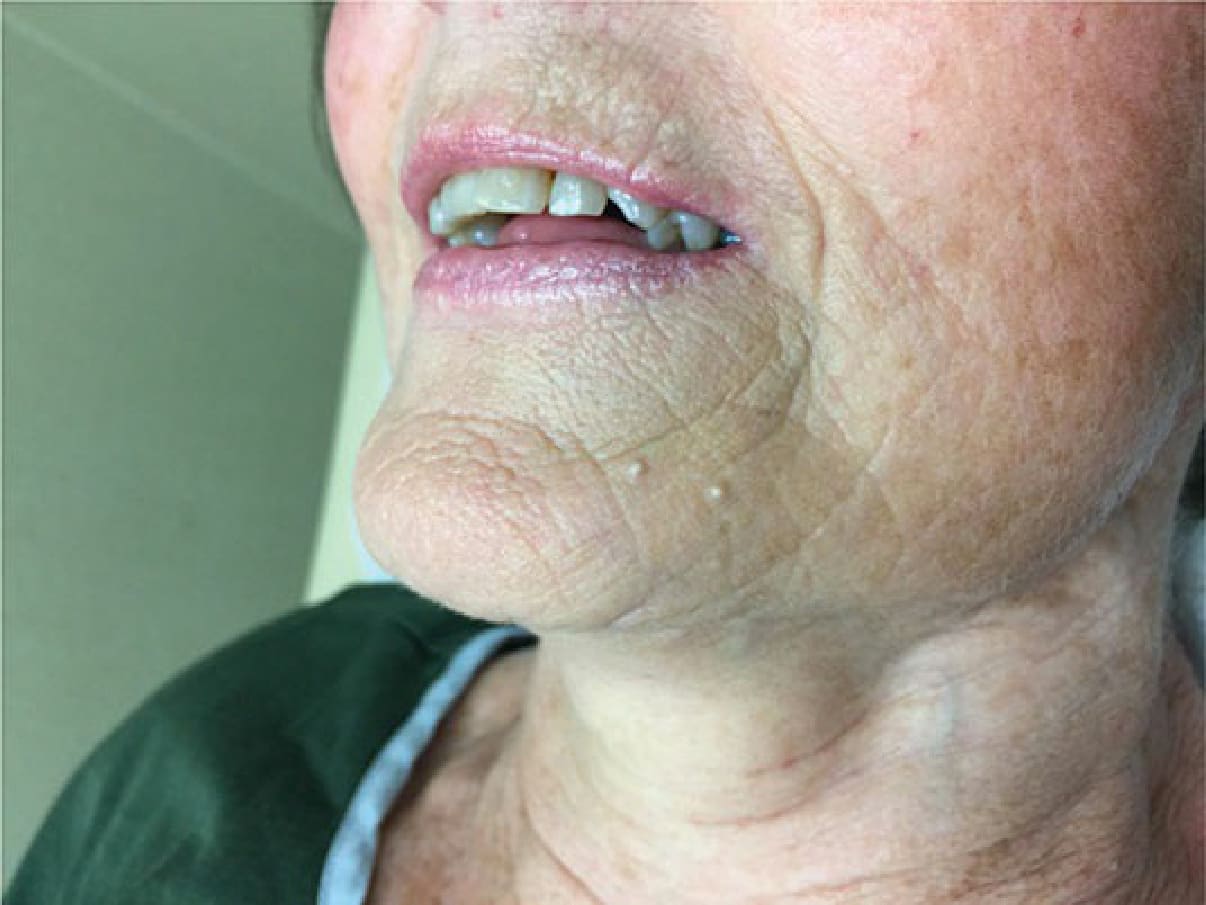
Figure 50-3. Primary milia are usually 1- to 3-mm white papules, most commonly occurring around the eyelids and central face.
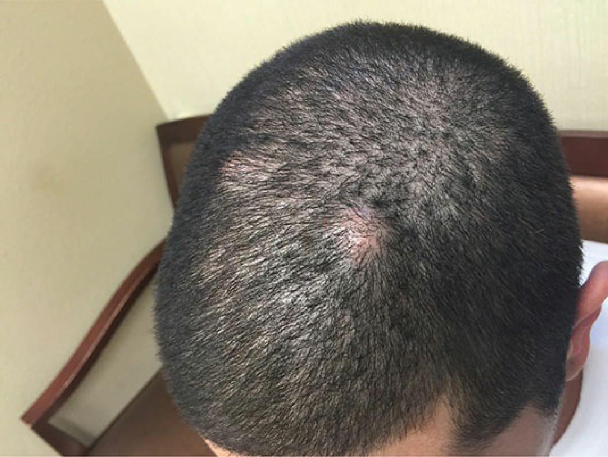
Figure 50-4. Unlike epidermal cysts, a punctum is usually not seen with trichilemmal cysts.
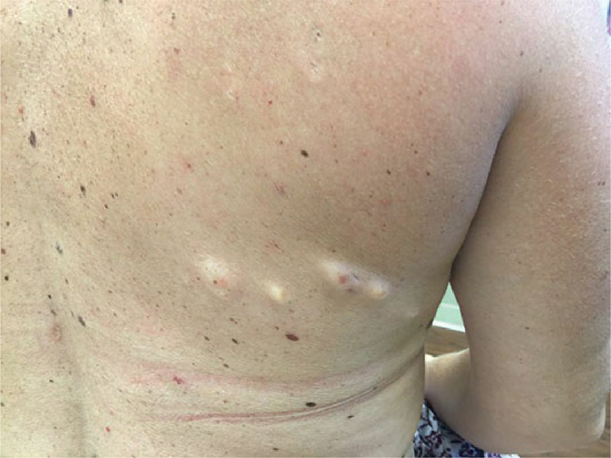
Figure 50-5. Compared to epidermal cysts, steatocystomas tend to be smaller, softer acneiform lesions (usually less than 1 cm) occurring on the chest and axillae.