LASER THERAPY
LASER THERAPY
Laser therapy represents an additional therapeutic option for keloids. Various lasers, both nonablative and ablative, have been studied with variable success. Nonablative lasers include pulsed-dye laser (PDL) and neodymium: yttrium-aluminum garnet (Nd:YAG), and ablative lasers include carbon dioxide and erbium:yttrium-aluminum garnet (Er:YAG).
Pulsed-Dye Laser PDL emits energy at wavelengths of 585- or 595-nm, targeting hemoglobin and oxyhemoglobin within red blood cells. This leads to selective photothermolysis of blood vessels, reducing vascularization of the keloid tissue and leading to tissue death and reduction in scar size.65 PDL has been studied extensively for the treatment of keloids, but has had mixed results as monotherapy. In one study, 16 patients were treated
with 585-nm flashlamp-pumped PDL and all patients demonstrated improvement in clinical appearance at 6 months.66 Another study evaluated the longer wavelength 595- nm PDL, and found superior results for patients with darker skin types.67 Other reports have shown minimal response or rapid recurrence following this treatment modality.68
A systematic review including eight randomized-controlled trials found PDL to be superior to conventional modalities in improving overall scar appearance, though this difference was not present when individual scar parameters were evaluated separately.69
The ideal fluence has not been defined, as no statistically significant difference was noted in one study between different fluences, and there was a trend toward better responses with lower fluences.67 General recommendations are for the fluence to be in the range of 4.5 to 7.5 J/cm2, depending on the spot size, with a higher fluence needed for smaller spot sizes.70
PDL can also be performed in conjunction with other therapies. A single-blind, randomized-controlled trial of 69 patients treated with either weekly IL TAC 10 mg/mL alone, weekly IL TAC with 5-FU (4 mg TAC with 45 mg 5-FU) or weekly IL TAC with 5-FU and 585-nm PDL for three sessions found the latter combination group to be most effective, with few side effects.71 PDL can also be used prior to IL injections as a means to facilitate injection by making the scar more edematous and more easily penetrable.72 Finally, PDL has also been used successfully post-shave excision in preventing recurrence.73
Adverse events of PDL include atrophic scarring, pigmentary changes, dermatitis, and purpura.74 Additionally, PDL has been reported to cause keloid formation when used for other indications.75
Nd:YAG Laser Nd:YAG laser emits light at a wavelength of 1064 nm, and therefore is able to penetrate deeper into the dermis. It is thought to suppress collagen synthesis by fibroblast inhibition.76 Though less studied than PDL for keloids, in small case series, Nd:YAG has been shown to improve the cosmetic appearance of keloids, including pigmentation, vascularity, and thickness.77,78 Side effects were often mild, and most commonly included transient post-treatment erythema. When compared to 595-nm PDL in a randomized split-scar trial of 20 patients with hypertrophic scars and keloids, both treatments produced statistically significant improvement as compared to baseline, though there was no significant difference between groups.79 More studies are needed to fully characterize the role of Nd:YAG in the treatment of keloids (Fig. 49-2).
Carbon Dioxide Laser Carbon dioxide (CO2) laser emits energy at a wavelength of 10,600 nm, which targets water within tissue, leading to vaporization and tissue destruction. Therefore, it functions as an ablative laser, and in treatment of keloids, causes necrosis of the keloidal tissue, remodeling of the scar, contraction, and ultimately, size reduction.80
CO2 laser has been used as monotherapy or in combination with IL steroids. As monotherapy, patients treated with CO2 laser with a 2 mm spot size and W to achieve a power density of 500 W/cm2 achieved successful results.81 When used in combination with IL steroids administered every 3 to 4 weeks, decreased recurrence rates were observed as compared to CO2 laser alone.82 Common side effects of CO2 laser include erythema and dyspigmentation.
Er:YAG Laser Er:YAG laser emits a 2940-nm wavelength that also targets water, but as it has a shorter wavelength than CO2 laser, does not penetrate as deep in the dermis, leading to reduced deep thermal damage.83 It has not been as extensively studied for the treatment of keloids as other lasers, but in a randomized controlled trial, Er:YAG was effective in improving the clinical appearance of hypertrophic scars with less side effects as compared with CO2 laser.84
CONCLUSIONS
Keloids represent an abnormal response to wound healing, and their management can be challenging. Approaches include IL therapies, such as steroids, 5-FU, verapamil, IFN, and bleomycin; cryosurgery, including IL cryotherapy; surgical excision; adjuvant
radiation therapy; and laser therapy. General surgical principles for tension reduction are of paramount importance in keloid surgery. Ideal management strategies vary based on lesion size and location, as well as patient preferences and motivation, and no one therapy is ideal in each situation. A recent treatment algorithm proposed beginning therapy with silicone gel or sheeting in combination with IL steroids (alone or in combination with 5-FU), followed by laser therapy; if refractory, the keloid may be treated with excision followed by other adjuvant therapies, including radiation. Further research is needed to clearly define the ideal approach to keloid management.
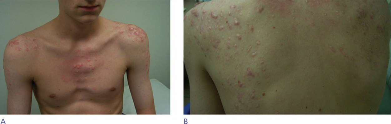
Figure 49-2. A young man with several keloidal papules on the chest, lateral arms, and upper back. The lesions are distributed in an acneiform distribution and were preceded by acne. In these cases, it is paramount to not only treat the bothersome keloidal lesions, but also treat the underlying acne, which is usually performed with more aggressive therapy if still active, in order to prevent new keloids.