EXCISION
EXCISION
For keloids refractory to IL therapies, surgical excision is often the next step in management. Surgical excision is typically performed in conjunction with adjuvant therapy to minimize recurrence rates, as excision alone can result in recurrence rates of 50% to 80%.39 Larger keloids and those present for shorter periods of time have a higher risk of recurrence.40 Several options for adjuvant therapies can be considered, with IL steroids representing a common approach. After excision, the wound edges can be injected with steroids, though in such cases, suture removal is often delayed to reduce the risk of wound dehiscence.41
In general, similar principles that are important for wound healing are helpful for keloid surgery, including minimizing local trauma, minimizing tension across the wound, and appropriate wound margin approximation.
General Surgical Principles Excision with primary closure is the main treatment modality performed for keloids that are amenable to this approach. This represents an appropriate option only if sufficient laxity of surrounding tissue is present so that the nascent wound is under minimal tension. If excessive tension is expected to be present, either serial staged surgical excisions or other surgical approaches (discussed below) should be employed. Excision of the keloid should be performed just deep to the junction of the keloid and normal, unaffected skin.42 Trauma to the deeper portion of the unaffected dermis should be minimized to theoretically reduce the risk of keloid recurrence. Some surgeons advocate keeping the incision just within the keloid margins and leaving a small rim of keloid tissue, though this approach has not been studied extensively.41 When deciding on
the appropriate excision margin, it may be helpful to note the vascularity of the tissue being incised, as keloidal tissue is less vascular than surrounding normal skin. Thus, once marginal bleeding occurs, the incision has entered into normal tissue. Additionally, a crunching sound is sometimes heard when incising the keloid proper, as the keloid is composed of fibrous tissue. The resolution of this sound signals suggests that unaffected skin has been entered.43
After excision, the tissue edges should be handled as atraumatically as possible, and only the minimal amount of undermining necessary to relieve wound tension should be performed.44 Potential sources of inflammation, such as trapped hair follicles, should be removed from the wound bed to minimize risk of recurrence.41
Suture Selection Suture selection is important in minimizing risk of recurrence, and in general, monofilament, synthetic suture may be preferable to braided suture given its decreased tissue reactivity, which may minimize the risk of microabscess formation and inflammation, thereby decreasing the risk of recurrence.41 Durkaya et al., evaluated the role of sutures in a randomized-controlled trial of 60 patients with sternotomy scars where the wounds were closed via a subcuticular suture that was either braided polyglycolic acid or monofilament polypropylene suture.45 Wounds closed with braided polyglycolic acid had significantly more hypertrophy than those closed with nonabsorbable monofilament polypropylene suture. Another study evaluated the risk of hypertrophic scar formation after breast reduction surgery, and found smaller and less reactive scars in patients whose wounds were closed with monofilament suture as compared to a braided suture.46 These small studies suggest that monofilament suture should be considered when excising keloids, though further research in this area is needed.
Tension Minimization Once suture material has been selected, the principle goal of suturing in prevention of keloid recurrence is minimization of tension. In addition to undermining and the selection of an appropriate closure technique for the given surgery, specific suturing techniques can be employed to minimize tension. Fascial plication sutures are sometimes employed to shift tension to the superficial and deep fascia, thereby reducing tension on the dermis and minimizing the need for dermal sutures.47 Similarly, utilizing the set-back dermal suture technique, rather than standard buried sutures or buried vertical mattress sutures, may similarly help shift injury away from the wound edges; this approach coupled with postoperative electron-beam radiation resulted in a 2-year recurrence rate of only 2.2%.48 A continuous intradermal suture has also been reported by placing nonabsorbable monofilament suture in both the deep dermis and superficial
dermis,49 which is then removed at the follow-up visit. Finally, a newer method for tension minimization after surgery is through use of a skin tension-offloading device. The Embrace Advanced Scar Therapy device was studied in a randomized-controlled split-scar trial of 65 adults status-post abdominoplasty, and found that mean visual analog scale score for embrace-treated scars was significantly improved compared with controls,50 though further study is needed.
Additional Surgical Approaches For keloids not amenable to primary closure, various surgical approaches have been developed. These include healing by secondary intention,51 healing with skin grafts,52
staged excisions, and surgical flaps.53 Healing by secondary intention has the disadvantage of a prolonged healing time, scar contracture and recurrence, while grafts involve the risk of donor site morbidity and color mismatch.53
The most important factor for a successful outcome after keloid surgery following an adjacent tissue transfer is to prevent flap necrosis by minimizing trauma to the flap and minimizing final wound tension.54 Another important principle in achieving a successful result is the use of adjuvant therapy post-surgery to minimize recurrence.
Several flaps have been specifically described for the treatment of earlobe keloids, where primary closure may lead to earlobe distortion and poor cosmesis. Adams and Gloster, reported use of a suprakeloidal flap for treatment of keloids.55 A similar technique, the keloid fillet flap, has also been described (Fig. 49-1).53 Another approach is through an X-shaped incision, which is marked on the surface of the keloid with the skin elevated from the surface of the keloid as four triangular flaps.54 The keloid tissue is surgically dissected and excised, and the defect is closed. Finally, a subcutaneous V-Y (island pedicle) flap may be effective for keloids on the posterior aspect of the ear.56
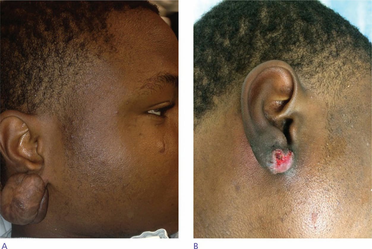
Figure 49-1. (A) Large keloid on the right earlobe. Previous treatment included intralesional steroids. The patient was treated with surgical excision using the keloid fillet flap, followed by adjuvant radiation. Postoperative visit showed slight dehiscence, which healed over the following weeks, and no recurrence was noted (B). Excision with adjuvant radiation is a good therapeutic technique when treating keloids located on the ears.