Technique
Technique
Local anesthetic drops are applied to the conjunctival fornix, followed by an injection
of local anesthetic containing epinephrine into the conjunctival lid margin, avoiding the marginal arcades.
Before the margin is closed, ensure the opposing tarsal plates have been cut perpendicular to the eyelid margin. This is essential to preserve the curve of the lid margin and avoid distortion or notching. The tarsal plate is recut if necessary.
The tarsal plates are reapposed with two sutures, a diagonal vertical suture and a horizontal inferior suture (Fig. 38-18). Each suture must take a deep bite of the tarsal plate. The superior diagonal suture is passed first into the right-hand tarsal plate 2 mm inferior to the lid margin and 2 mm inside to the cut edge. The tip exits just deep to the conjunctiva at the uppermost aspect of the tarsal plate. This diagonal pass is most easily achieved by grasping the tarsal plate horizontally at its midpoint. The second pass of the superior diagonal suture is into the left-hand tarsal plate. This is most easily achieved by backhanding the suture, grasping the tarsal plate vertically and entering the tarsal plate just deep to the conjunctiva at the uppermost point of the tarsal plate and passing to exit the anterior surface of the tarsal plate 2 mm inferior and 2 mm inside the cut edge. This superior suture is clipped and neither tied nor cut to allow easier access for the next suture. The second horizontal suture is then passed through the tarsus horizontally at the inferior border of the tarsal plate with a generous bite of each tarsal plate, remaining deep to the conjunctiva. This can be tied and cut. The clip from the superior diagonal suture is released and the suture tied and cut. The apposed edges of the lid margin should be in perfect apposition. The closure of the posterior lamella should now be complete. The anterior lamella is then closed in a layered manner with deep sutures closing the orbicularis and skin sutures beginning at the subciliary line. Topical antibiotic ointment is applied to the eye and sutures daily until the skin sutures are removed in 6 days. Ice packs are applied for comfort and to reduce swelling.
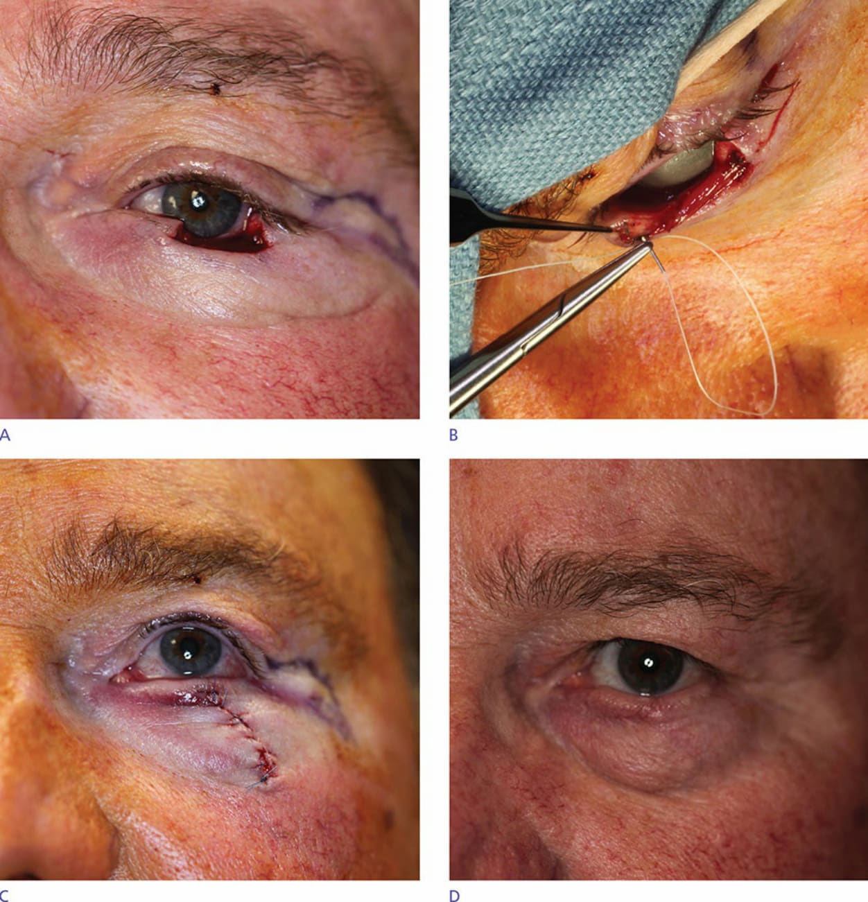
Figure 38-18. Primary lid repair: Diagonal tarsal suture sine marginal sutures. (A,B) Full-thickness defect involving medial the lid margin. (C) Primary lid closure postoperatively, robustly approximated, and accurately aligned without need for marginal sutures. (D) Lid snug against the globe without notching 3 months postoperatively.