Excessive Granulation Tissue
Excessive Granulation Tissue
Excessive granulation tissue is considered a type of abnormal wound healing and is often seen in wound healing by second intention.150 Associated factors include wound site, prolonged inflammation, an imbalance in matrix metalloproteinases, and excessive angiogenesis. While a modest amount of granulation tissue is considered favorable in wound healing by second intention, too much granulation tissue inhibits wound healing by preventing fibroblast proliferation. Granulation tissue appears beefy red and friable, and may extend above the level of normal skin. Management of excessive granulation tissue includes destruction with silver nitrate, laser ablation, cautery, curettage, or shave removal of the tissue. Medium- to high-potency topical corticosteroids have also been used with success.150,151
Scarring Scarring after skin cancer surgery can profoundly affect psychosocial functioning, especially when scars are located on the head and neck.152 For many patients, the success of a surgical procedure is often tied to the aesthetic appearance of the final scar. Surgical scars tend to have a variable presentation, and may have multiple features that must be addressed for optimal scar reduction. Scars may be characterized by abnormal erythema or pigmentation, firmness, visibility, contour irregularity, and other negative factors. Additionally, the position, location, age of the scar, and skin type of the patient should be considered. For many surgical scars, time will resolve many of the adverse
features. Laser therapy with multiple different energy modalities has been shown to improve scar features.153,154 Dermabrasion is another method for treating surgical scars, and is particularly useful on sebaceous nasal skin.155 Surgical scar revision may be attempted if the scar is not exhibiting favorable characteristics for long term. Multiple techniques may be utilized, including scar excision with linear closure, Z-plasty, Wplasty, and geometric broken line closure.155 Surgical scar revision techniques are addressed in detail in Chapter 35.
Hypertrophic/Keloid Scars. Hypertrophic scars and keloids are two forms of abnormal wound healing characterized by local fibroblast proliferation and excessive collagen production in response to cutaneous injury. Hypertrophic scars tend to remain confined to the scar line, whereas keloids extend beyond the margin of the scar. Hypertrophic scars often arise earlier than keloids, usually within 4 weeks postoperatively. Keloids may develop months to a year after trauma or surgery.156 Hypertrophic scars can regress without intervention, whereas keloids do not, and indeed frequently recur after surgical removal. Keloids are more common on the chest and back of darker-skinned patients, and are often also seen at the sites of wounds healing by second intention.157 In contrast, spread scars tend to be more atrophic, with thin, fragile tissue centrally. Spread scars occur in areas under high tension, high use (shoulders, chest, back), and in the context of infection or dehiscence.
The first-line treatment for hypertrophic scars and keloids is intralesional steroid injections.158 While this approach is considered largely effective, it may be associated with cutaneous atrophy, pigmentary alteration, and telangiectasias.159 Re-excision with the addition of pressure therapy, postoperative radiation, and intralesional steroid injections has also been performed with success for keloids,160,161 and one study has demonstrated an effective combination of re-excision and closure with the set-back suture coupled with postoperative radiation.162 Finally, silicone gel sheeting, 585- or 595-nm pulsed-dye laser, cryotherapy, intralesional 5-fluorouracil (5-FU), interferon alfa-2b injections, fractional nonablative lasers, and ablative laser treatment may also be of benefit for keloids.163–168
Suture Track Marks. Track marks are often seen when epidermal sutures are left in place longer than necessary or are placed too tightly (Fig. 36-13).104 Postoperative edema tends to exacerbate track marks due to increased tension on epidermal sutures. To prevent track marks, sutures should be removed as early as possible, wound tension should be maximally decreased by performing adequate undermining, buried dermal or subcutaneous sutures should be used for tension reduction, and sutures of appropriate size should be used for a given anatomic location. A recent randomized controlled trial found the set-back suture to be superior to the buried vertical mattress suture for postoperative scar cosmesis.148 Tissue adhesives or adhesive strips may also be used in
place of epidermal sutures in order to prevent track marks as long as dermal sutures have been placed to reduce tension.169
Trapdoor Phenomenon The trapdoor phenomenon, or pin-cushioning, is a contour irregularity due to scar elevation above the surrounding skin surface. This abnormal scarring is most frequently seen in flaps on the nose and upper cutaneous lip (Fig. 36-14). Irregular flap contour may be due to several factors, including lymphatic or venous obstruction, postoperative ischemia, resolving hematoma, scar hypertrophy, excessive subcutaneous fat or flap tissue, and scar contracture. The deformity typically appears within 3 weeks to 6 months after surgery and can be prevented with optimal flap design, wide undermining of the recipient site, squaring of the corners (versus circular trimming), appropriately thinning the flap, and the judicious use of dermal sutures to re-approximate muscle and dermis. Potential treatments include surgical revision by lifting and thinning of the flap, intralesional steroid injection, and dermabrasion. Over time, improvement may be seen without intervention.170,171
Free Margin Retraction Free margins are anatomic areas where the skin surface remains unattached to the surrounding tissue. Common free margins on the face are the eyelid, helical rim of the ear, alar rim of the nose, and the vermilion lip. Because they offer little resistance to tension created by surgical reconstruction or scar contracture, free margins are vulnerable to distortion (Fig. 36-15).
Disruption of these natural contours is both aesthetically displeasing and functionally significant. Ectropion may result in difficulty closing the eyelids with subsequent dry eyes and corneal irritation, alar rim retraction can cause nasal valve obstruction, and eclabium can lead to challenges when creating a tight oral seal.
To prevent these complications, surgical closures must be designed to minimize tension on the free margin; this is often achieved by orienting closure tension vectors perpendicular to the free margins to avoid “pulling” the free margin out of position.172 Cartilage grafts may be used to prevent alar rim retraction, and Frost sutures may be used to prevent ectropion in the immediate postoperative period, though they cannot compensate for poorly designed closures.173 Caution should be used when using second-intention healing or when placing skin grafts in locations near a free margin, as clinically significant contraction may occur. Treatment of free margin retraction includes intralesional steroids and scar revision.
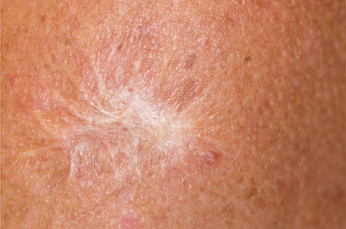
Figure 36-13. Spread surgical scar.
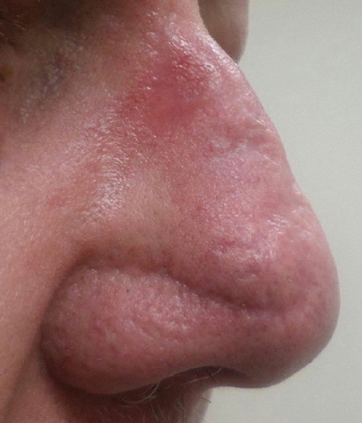
Figure 36-14. Pin-cushioning or trapdoor deformity of transposition flap on the nose.
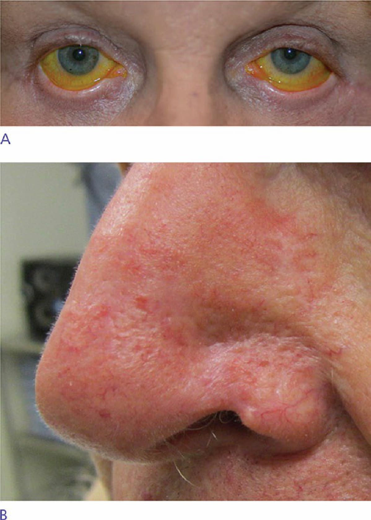
Figure 36-15. Contracture of a free margin resulting in (A) ectropion after lower blepharoplasty and (B) alar retraction after interpolated flap necrosis.