Sensory Nerves
Sensory Nerves
Cutaneous nerves are commonly transected during dermatologic surgery. As a result, patients frequently experience paresthesias at the procedure site. Given that most areas on the skin have diffuse sensory innervation, and sensory nerves often regenerate, there are rarely significant permanent deficits from cutaneous surgery. Patients should be counseled preoperatively about risk of paresthesias at the surgical site, and that normal sensation usually returns within 18 months. Occasionally, complete sensation never returns. Areas most prone to sensory nerve injury and appreciable deficits are the digits, forehead, and scalp. In particular, procedures that require deep tissue removal or dissection risk permanent damage to the branches of the trigeminal nerve on the face, with subsequent sensory loss.
Motor Nerves Injury to motor nerves can result in more severe consequences than damage to sensory nerves. Though rare, damage to motor nerves can cause significant functional impairment. While many of the muscles of the head and neck have significant redundancy in innervation, there are specific motor nerve “danger zones” that are most at risk during dermatologic surgery. Overall, the risk of motor nerve damage in dermatologic surgery is very low, but patients need to be counseled preoperatively when operating in high-risk locations. Surgeons also need to be aware that significant individual variability exists in the anatomic course of nerves. Additionally, patients with tissue atrophy, such as seen with normal aging or HIV lipoatrophy, will have motor nerves coursing in a more superficial plane. If motor nerve damage occurs, it may be temporary (neuropraxia) or permanent. It is prudent to consult with the appropriate specialty (generally neurology or otolaryngology) early to optimize management after motor nerve damage. The nerves at the greatest risk for injury during cutaneous surgery are the temporal and marginal mandibular branches of the facial nerve and the spinal accessory nerve.44
The temporal branch of the facial nerve is the most commonly injured nerve in facial
surgery. When damaged, it will lead to ipsilateral brow ptosis secondary to paralysis of the frontalis muscle (Fig. 36-9).139 The nerve branches off the facial nerve trunk within the parotid gland and courses superficially across the zygomatic arch within the innominate fascia (a plane deep to the SMAS and superficial temporal fascia), and crosses the temple with little overlying subcutaneous fat.140 If nerve damage is permanent, this complication can be addressed with a direct or indirect brow lift. Over time, patients may develop a compensatory brow elevation on the contralateral side of injury.139
The marginal mandibular branch of the facial nerve traverses the mandible near the facial artery and vein, covered only by skin and thin platysma muscle. While the nerve is normally located approximately 1 to 2 cm below the mandible, in individuals with lax or atrophic tissues (as seen with aging), the branches can be as low as 3 to 4 cm below the mandible.141 This nerve innervates the lip depressors, and damage presents clinically as asymmetric facial expressions and mouth function compromise. This asymmetry and lip imbalance are readily noticeable during opening of the mouth (Fig. 36-10).142 The marginal mandibular nerve is at risk during liposuction of the jowls and neck dissections, as well as after deoxycholic acid injections for submental fat.143,144 The buccal and zygomatic branches are deeper, with more collateral innervation, and are at less risk of damage when compared to the other branches of the facial nerve.
Finally, damage to the spinal accessory nerve results in a winged scapula due to trapezius muscle palsy. This palsy results in dropping of the shoulder girdle inferiorly
and laterally along with flaring of the wing of the scapula and loss of abduction of the arm.44 The spinal accessory nerve exits behind the sternocleidomastoid (SCM) muscle at the junction of the upper and middle third of the SCM, then courses across the posterior triangle of the neck. It primarily innervates the trapezius and partially innervates the SCM. Inadvertent transection can occur during procedures in the posterior triangle of the neck, including radical neck dissection, lymph node dissection, and extensive cyst or tumor resection.
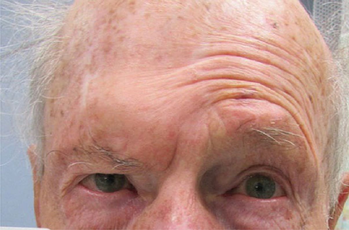
Figure 36-9. Injury to the temporal branch of the facial nerve resulting in the inability to raise the right brow due to frontalis muscle paralysis.
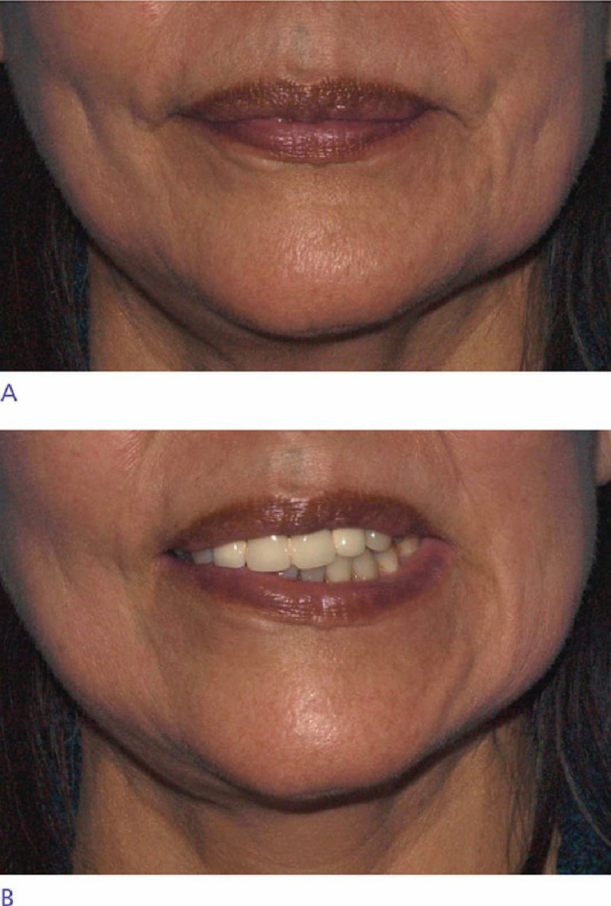
Figure 36-10. Marginal mandibular nerve paralysis after neck dissection, with injury on the right side: (A) At rest; (B) smiling showing compensation on the left side.