Skin Necrosis
Skin Necrosis
Tissue ischemia is a result of decreased vascular perfusion that does not allow for adequate tissue perfusion. Ischemia can eventually result in tissue necrosis or death.99
Black, densely adherent eschar represents tissue necrosis (Fig. 36-7).6 Skin necrosis is a rare complication seen in cutaneous surgery. Necrosis is often seen as a result of patient risk factors, anatomic risk factors, poor closure design (e.g., excessive tension), postoperative bleeding, or wound infection. Patient risk factors include anticoagulant use, hepatic or renal insufficiency, excessive alcohol consumption, and smoking (heavy smokers develop necrosis three times more frequently than never smokers).100,101 Tobacco use decreases cutaneous blood flow via nicotine (causes vasoconstriction) and carbon monoxide (impairs cutaneous oxygenation). It is recommended that patients decrease their smoking to <1 pack per day for 2 days prior to surgery and for 1 week postoperatively to minimize complications.6
Proper surgical technique also plays a major role in preventing ischemia and necrosis. Delicate tissue handling is important. The surgeon should aim to avoid macerating tissue edges; this can be achieved by using skin hooks or toothed forceps rather than serrated forceps.6,99 When undermining, the surgeon should avoid disrupting the dermal plexus by undermining at the appropriate depth.102 Strangulation of tissue with sutures can also lead to tissue ischemia. Elastic sutures may have a lower risk of necrosis because the suture material can expand with tissue edema.103 The tip stitch, or half-buried mattress suture, minimizes compression on the subdermal vessels in order to minimize ischemic necrosis of vulnerable flap tips.104 Mattress sutures, in contrast, may increase the risk of necrosis. Finally, prevention of hematoma, as noted above, may also prevent tissue ischemia and necrosis.
From the standpoint of closure design, high-tension closures should be avoided in order to minimize the risk of tissue ischemia.105 Techniques to minimize closure tension include appropriate undermining, using plication sutures and/or dermal and
subcutaneous sutures to decrease tension on the epidermal edge,106 and using relaxation incisions or Z-plasties. Flaps and grafts have an increased risk of tissue ischemia compared to primary closures.105 In flaps, the vascularity is based only on the pedicle supplying the tissue. The length of the pedicle correlates with the risk of flap necrosis. One study examining dermatologic surgery complications prospectively found that 4 of 241 patients (1.7%) of flap repair cases had evidence of partial flap necrosis; no complete flap necrosis occurred. The authors also found no significant association between anatomic site and flap necrosis. Graft necrosis tends to occur more frequently. In the same study, partial graft necrosis occurred in 13 of 152 patients (8.6%). Average graft loss was 50% of the total graft area.59
The first signs of ischemia and necrosis may be seen intraoperatively. In the event of arterial insufficiency, the tissue tends to be dusky or pale and cool to the touch. If venous congestion is present, the tissue is typically dark purple and bleeds readily if pricked. In both of these settings, the surgeon should redesign or modify the closure to maximize blood flow. In the postoperative period, arterial ischemia can be reversed by early intervention, as the tissue is capable of surviving up to 13 hours. In venous congestion, the tissue progresses to necrosis much more rapidly. Serial pinpricks can be performed in the postoperative period to mitigate venous congestion. Postoperatively, tissue necrosis appears as a dusky eschar with tissue sloughing. Nonviable tissue should not be debrided given the risk of damaging underlying viable tissue. The wound should be kept moist and occluded, and patients should be counseled on the expected clinical course, including tissue sloughing and odor. Once the necrotic tissue has completely sloughed off, healing by second intention or surgical intervention can be considered. No surgical closure should be attempted with devitalized tissue still present in the wound bed.
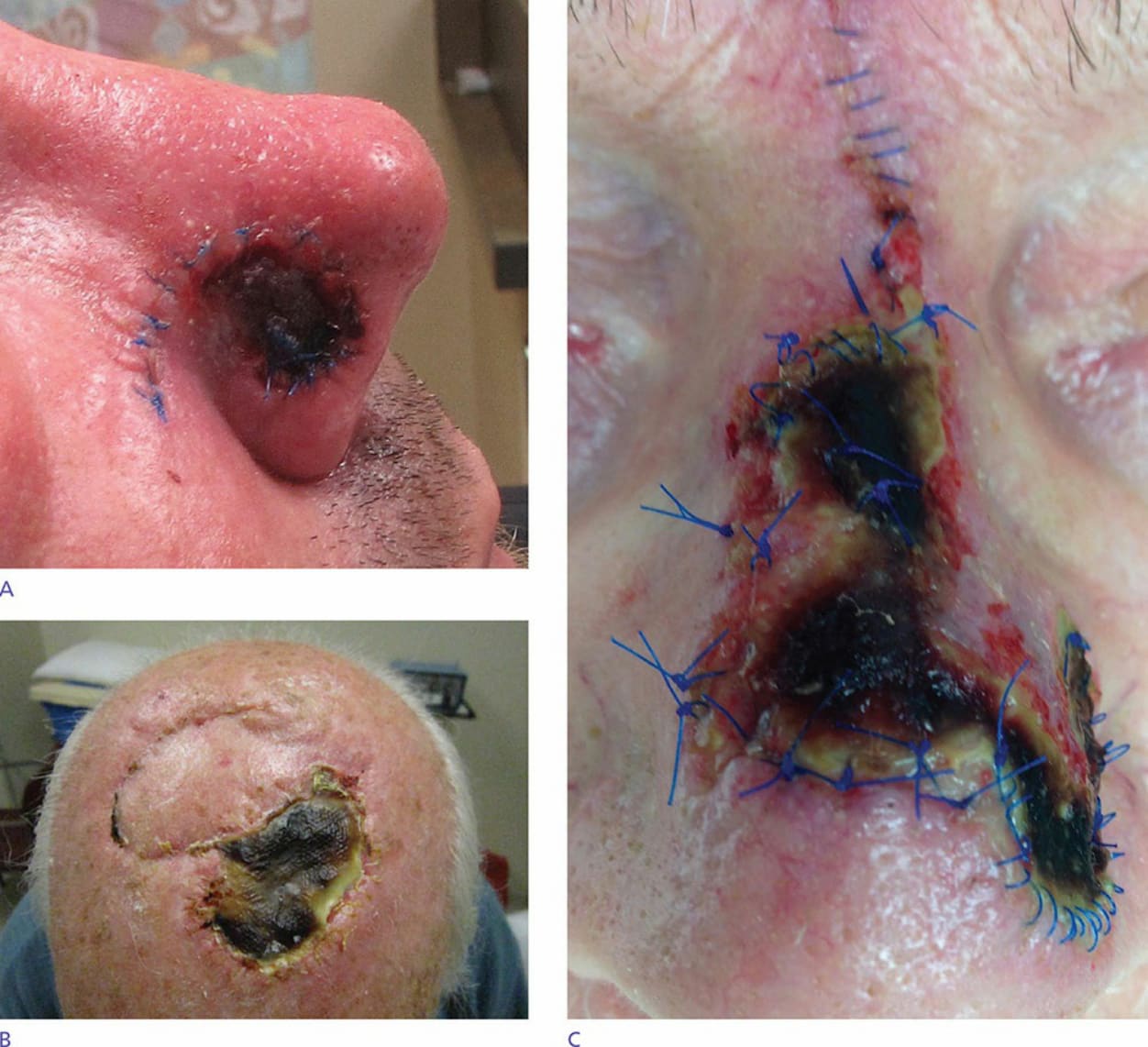
Figure 36-7. (A) Impending necrosis of rotation flap. (B) Tissue necrosis of full-thickness skin graft. (C) Tissue necrosis of transposition flap.