Complications Associated With Electrosurgery
Complications Associated With Electrosurgery
Cardiac Implantable Electronic Devices In North America alone, there are more than 250,000 new cardiac devices implanted each year.72 As older patients are at higher risk for both skin cancers and cardiac conditions requiring implantable electronic devices (IEDs), understanding the potential implications of electrosurgery is prudent for the dermatologic surgeon.73 Electromagnetic interference (EMI) from electrosurgical devices is relatively rare, as electrosurgery in office-based settings generates relatively low electromagnetic fields. In 2000, a survey of 166 Mohs surgeons revealed a low rate of complications due to electrosurgery (0.8 cases in 100 years of surgical practice) with no significant morbidity or mortality.74 Complications included skipped beats (eight patients), reprogramming of a pacemaker (six patients), firing of an ICD (four patients), asystole (three patients), bradycardia (two patients), depleted battery life of a pacemaker (one patient), and an unspecified tachyarrhythmia (one patient). Bipolar forceps were not associated with any complications.
Cardiac pacemakers are electronic devices consisting of a pulse generator and leads which provide electrical stimulation to cause cardiac contraction if the intrinsic heart rhythm is absent or slowed.75 In general, pacemaker patients are classified as dependent or independent. Pacemaker-dependent patients have inadequate or even absent intrinsic rhythm, and therefore can suffer significant symptoms or even cardiac arrest after cessation of pacing.76 As a result, patients who are pacemaker dependent are at higher risk of complications from EMI during electrosurgery. Implanted cardioverterdefibrillators (ICDs) can detect and terminate life-threatening tachyarrhythmias via high-energy shocks, and pace bradyarrhythmias. If EMI is erroneously identified as a life-threatening arrhythmia, a shock may be delivered or the ICD may respond by inhibiting cardioversion or pacing.77 ICDs are more sensitive to EMI than pacemakers. Additionally, with poorly grounded or nonisolated electrosurgery, an implantable cardiac device can work as an indifferent plate and cause myocardial burn or arrhythmia because the delivered energy is localized to a small area.74 Safeguards against EMI are under continuous development and include insulated coatings to limit the distance current must flow between electrodes and protective algorithms and filters that limit nonphysiologic interference.72,78 As a result of the protective circuitry of modern pacemakers, complications from EMI are currently quite rare.72
As office-based monopolar electrosurgical units such as hyfrecators are low powered compared to their hospital operating room counterparts, a grounding pad is not consistently required.78 Without a grounding pad, current disperses throughout the body and results in relatively safe means of obtaining hemostasis in most patients. However,
since the current travels to a distant site, there is a potential risk of EMI with cardiac IEDs. In bipolar electrosurgery, current travels through a two-electrode instrument from one electrode, through tissue, to a second adjacent electrode, completing an electrical circuit. A grounding pad is not required and electrical energy capable of interfering with an IED is minimized.78 A recent in vitro study observed that monopolar electrosurgical units did not interfere with defibrillators, and affected pacemakers only when used in close proximity to the device.79 It was concluded that these devices are safe to use in patients with defibrillators at any distance and within 2 inches of pacemakers. However, conservative guidelines advise that electrosurgery should be avoided within 15 cm of a cardiac device without consultation from an electrophysiologist.78 The safest forms of cautery in patients with cardiac IEDs are electrocautery (heat cautery) and electrosurgery with bipolar forceps (Fig. 36-3).
A magnet placed over a pacemaker will switch most demand pacemakers to an asynchronous mode.72,80 In this mode, the heart is paced at a fixed rate regardless of the patient’s underlying rhythm.78 This method has been used to protect patients from the inhibition of the pacemaker by EMI. When the magnet is removed, the device should revert to previous programmed pacing. Rare case reports of pacemaker malfunction have been associated with magnet use.35,77 For ICDs, tachycardia detection can be disabled by magnet application without having an effect on pace mode or rate. Most ICDs will revert to prior arrhythmia detection upon the removal of the magnet. An
important feature unique to ICDs is that magnet response will not affect ICD antibradycardia pacing functions.80 Unlike pacemakers, the pacemaker function of an ICD will not be rendered asynchronous. Thus, if a patient is dependent on the intrinsic antibradycardia pacing of their ICD, they will be potentially vulnerable to bradyarrhythmias from electrosurgery-induced EMI inhibiting the pacing function.80
Surgeons should obtain direction from a cardiologist or the patient’s pacemaker/ICD clinic prior to the use of magnets due to the low but potentially significant risks.
Noncardiac Implanted Electrical Devices Noncardiac IEDs include deep brain stimulators, spinal cord stimulators, vagal and phrenic nerve stimulators, gastric stimulators, and cochlear implants. These IEDs can also develop complications related to EMI.78 To limit the risk of EMI, many of these devices can be turned off with an external remote control. However, some devices cannot be inactivated due to functional or medical reasons. Electrosurgery can lead to electrode heating and tissue injury around the stimulator, as well as paresthesias and electrical shocks, even if the device is deactivated.72 As with cardiac devices, the risk is the greatest with monopolar electrosurgical devices. If a noncardiac IED cannot be deactivated, or the surgeon is working close to the electrodes, the physician managing the device should be consulted.
Burns and Fires Approximately 50 to 100 surgical fires occur in the United States each year, with the majority involving either electrosurgery or laser devices.81,82 Of these cases, one to two per year result in death.82,83 In dermatology specifically, electrosurgical pencil tips for coagulation are a common ignition source.82 Dermatologists also routinely use flammable and combustible chemicals, such as surgical dressings, drapes, and certain cleansers (i.e., isopropyl alcohol).17 Petrolatum has not been found to be flammable and can be safely used in a surgical field.84 Supplemental oxygen has been found to be a contributing factor in 74% of all surgical fires.81 Because 90% of surgical fires are caused by monopolar electrosurgical units and laser devices, bipolar electrosurgery is the preferred method of coagulation by the Emergency Care Research Institute as a means to prevent surgical fires in situations where oxygen supplementation is required.81 In the majority of dermatologic surgery procedures, patients’ oxygen can be turned off while using electrosurgery without adverse effects. All surgical personnel should be trained in fire safety procedures, including initiating a “Code Red” and knowing the location of fire extinguishers and fire alarms.
Burns can also be a major complication secondary to electrosurgery.82 In one survey of otolaryngologists, 324 complications from electrosurgical instruments were reported out of 99,664 cases performed in 1 year. These complications included 219
unanticipated direct burns, 48 burns secondary to current flow through a metallic retractor or instrument, 13 grounding pad burns, and 11 fires.85 Grounding pad burns typically occur due to a pad with dry gel, an improperly sized pad, moisture under the pad, or improperly positioning the pad. Single-use grounding pads should be placed on clean, dry skin overlying a well-perfused large muscle ipsilateral and close to the surgical site.82
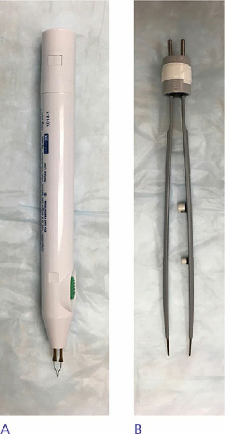
Figure 36-3. (A) Electrocautery and (B) bipolar forceps. These two methods of cautery are the safest in patients with implantable electrical cardiac devices.