7).40
7).40
Subcision may be performed to achieve ideal contour in cases of trapdoor deformity. An incision is made at the distal edge of the elevated flap (Fig. 35-8). Undersurface debulking is performed followed by re-approximation of the flap. Concavity revision may be more challenging, though a free cartilage graft may be used to raise the nasal tip to improve the appearance of a failed full-thickness skin graft (Fig. 35-9).
following failure of skin graft. (B) Free cartilage graft was obtained from the antihelix. (C) Graft was placed after incision and elevation of site. (D) Contour improvement at 2 months.
CONCLUSIONS
Scars are an inevitable consequence of cutaneous surgery. The goal of surgical scar revision is improved functionality and cosmesis. Consideration of the type of scar, location of the scar, quality and texture of the skin, RSTLs, and other considerations are all essential in planning an ideal revision. Notably, regard for tension on the healing wound is of paramount importance. Use of nonsurgical and surgical techniques often leads to the most ideal revision. Meticulous planning, including an appreciation of the timeline for various scar revision techniques, is also essential. By appreciating the ever-expanding armamentarium of surgical scar revision techniques, the surgeon may better care for their patients and produce optimal outcomes.
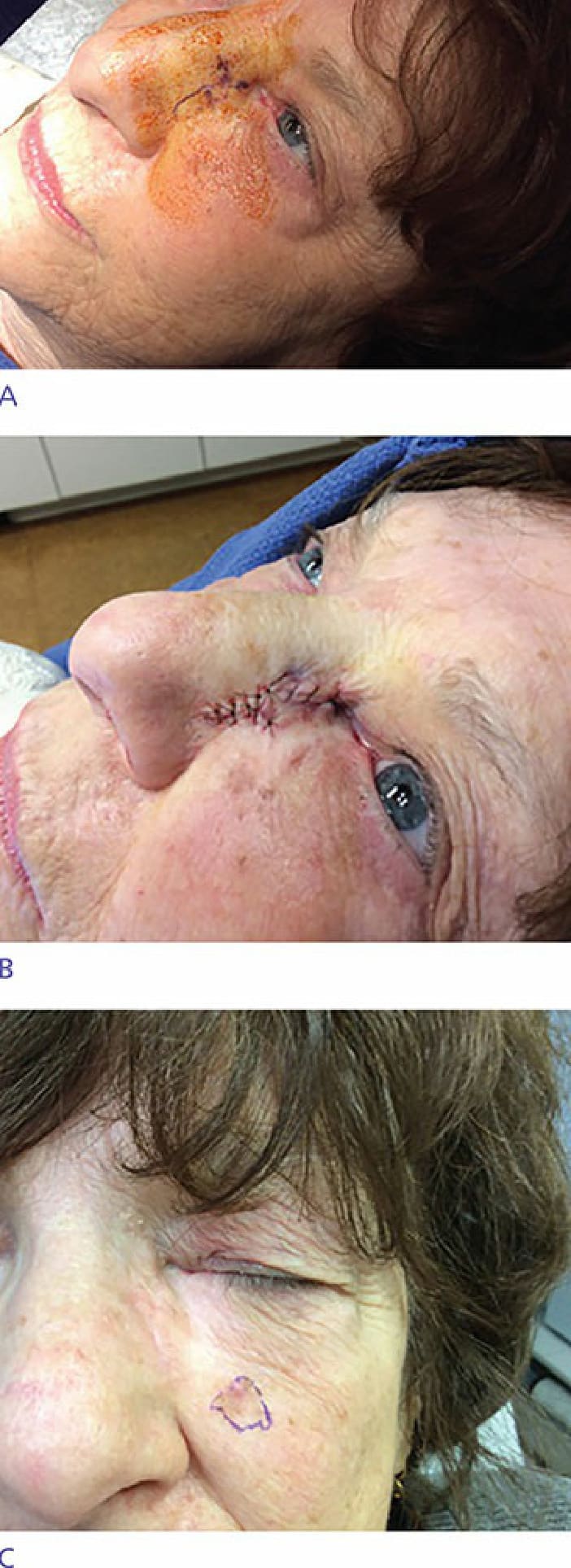
Figure 35-7. Rotation flap with back cut and periosteal anchoring suture to correct medial lower lid ectropion. (A) Preoperative view with ectropion resulting in tearing. (B) Immediate postoperative view. (C) Patient achieves normal eye closure with resolution of tearing.
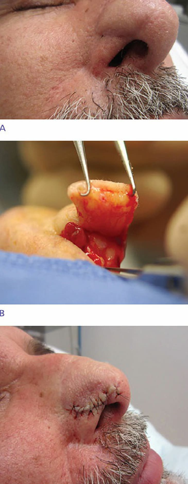
Figure 35-8. Subcision technique for revision of trap door deformity. (A) Trap door deformity at distal portion of paramedian forehead flap. (B) Area is incised, elevated, and deep aspect of the bulky area is thinned. (C) Flap is reapproximated after placement of buried absorbable suture to recreate the alar crease.
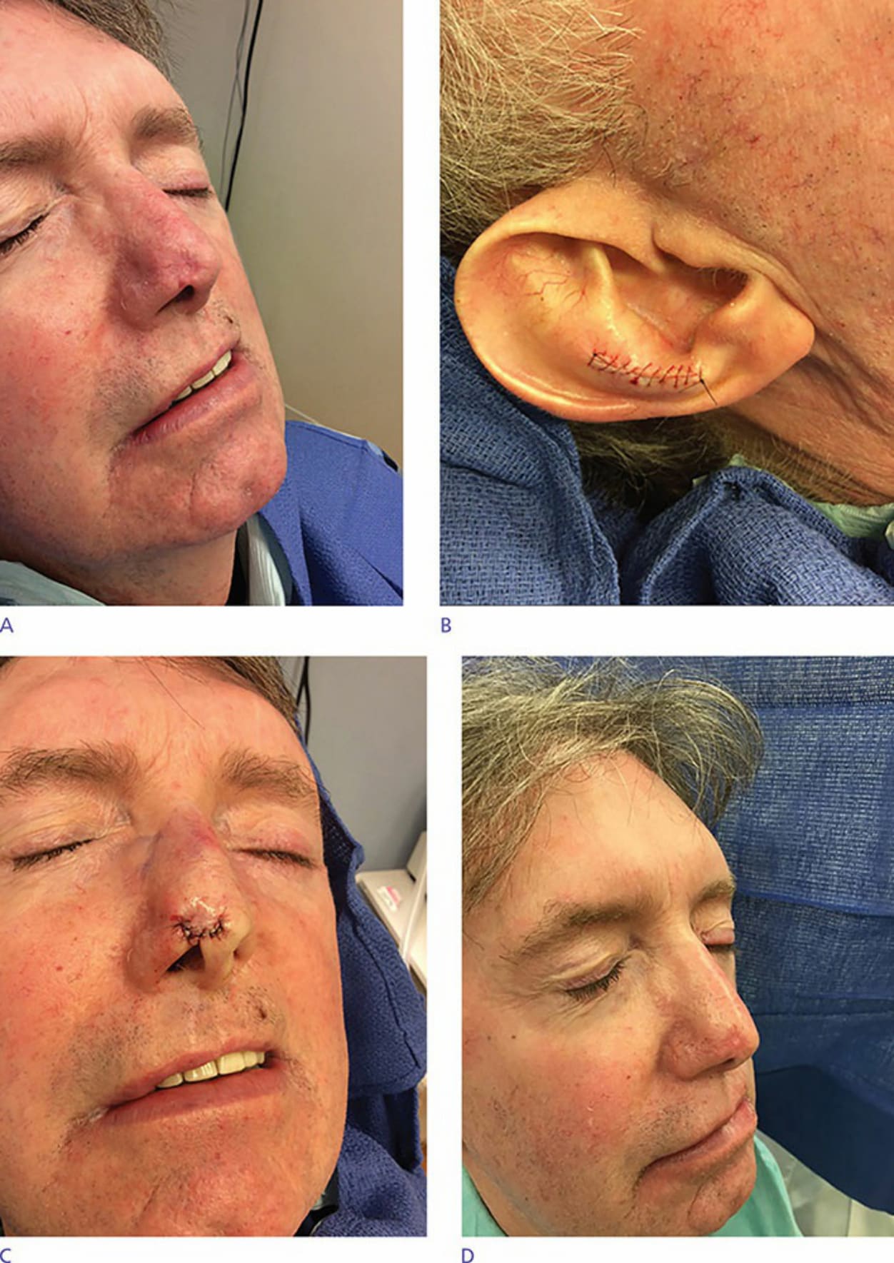
Figure 35-9. Placement of a free cartilage graft to restore contour. (A) Preoperative view after referral for revision