ANATOMY
ANATOMY
The nail unit is composed of nail plate, nail matrix, nail bed, hyponychium, nail folds, nerves, and blood vessels (Figs. 34-1 and 34-2). The latter derive from branches of the common volar digital arteries that have arterial anastomoses over the dorsal surface of the distal phalanx (Fig. 34-3). Innervation to the nail unit is supplied by sensory nerves that travel along the lateral aspects of the digits in close proximity to the aforementioned arteries. The perionychium (tissues surrounding the nail plate) is supplied by the common volar digital nerves which branch dorsally distal to the distal interphalangeal joint.1 Venous drainage of the perionychium coalesces laterally and dorsally, drains in a random fashion over the dorsum of the digit, and progresses toward anastomoses at the level of the distal interphalangeal joint. Lymphatics of the nail unit are parallel to the venous drainage, and are most dense at the hyponychium.
A comprehensive physical examination is critical in localizing the origin of nail unit pathology. The proximal nail matrix gives rise to the outer surface of the nail plate, while the distal nail matrix produces the undersurface of the nail plate.
Examination of the free edge of the nail allows the clinician to determine whether the disorder originates in the proximal matrix (Fig. 34-4) or in the distal matrix (Fig. 34-5).
indicates that the lesion of interest is located in the distal matrix.
After examination of the digit and identification of the source of nail pathology, the surgeon may discuss with the patient the various surgical approaches that are relevant for the pathology.
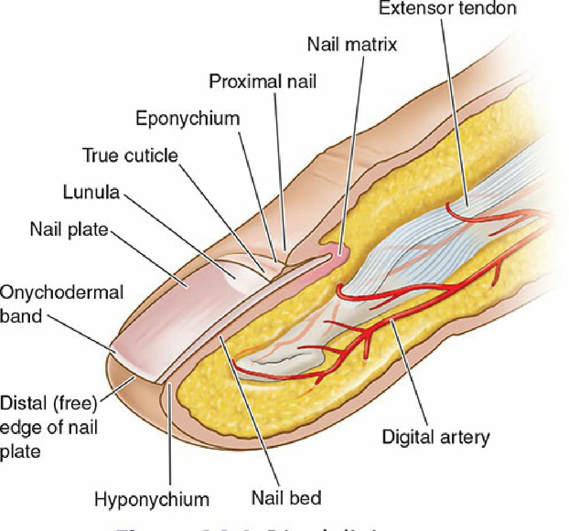
Figure 34-1. Distal digit anatomy.
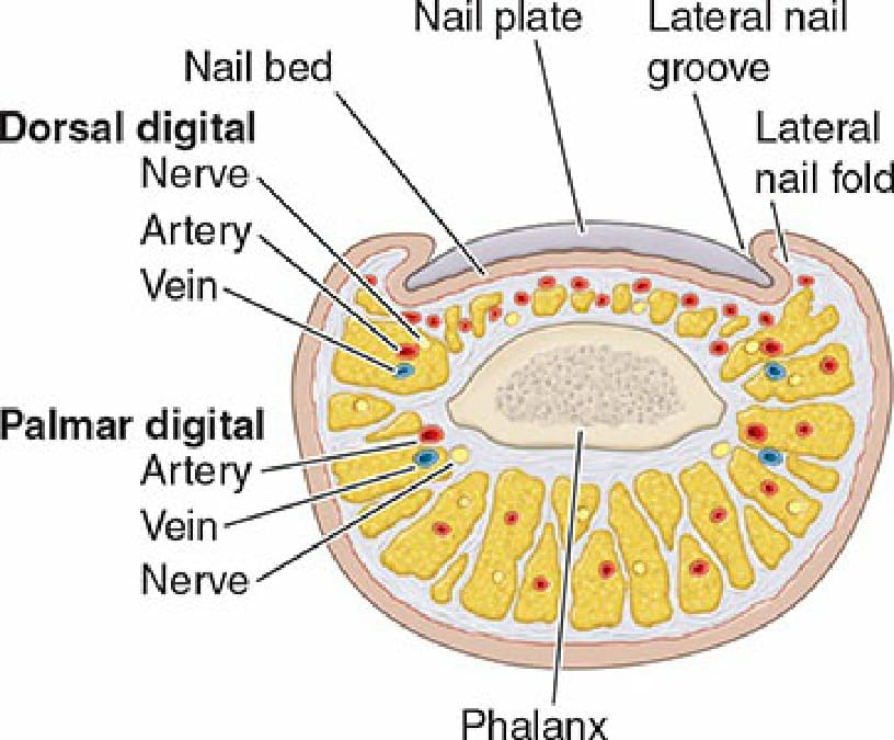
Figure 34-2. Transverse section of anatomy through the distal digit.
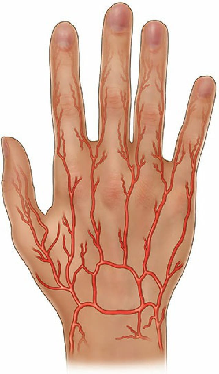
Figure 34-3. Dorsal hand arterial supply.
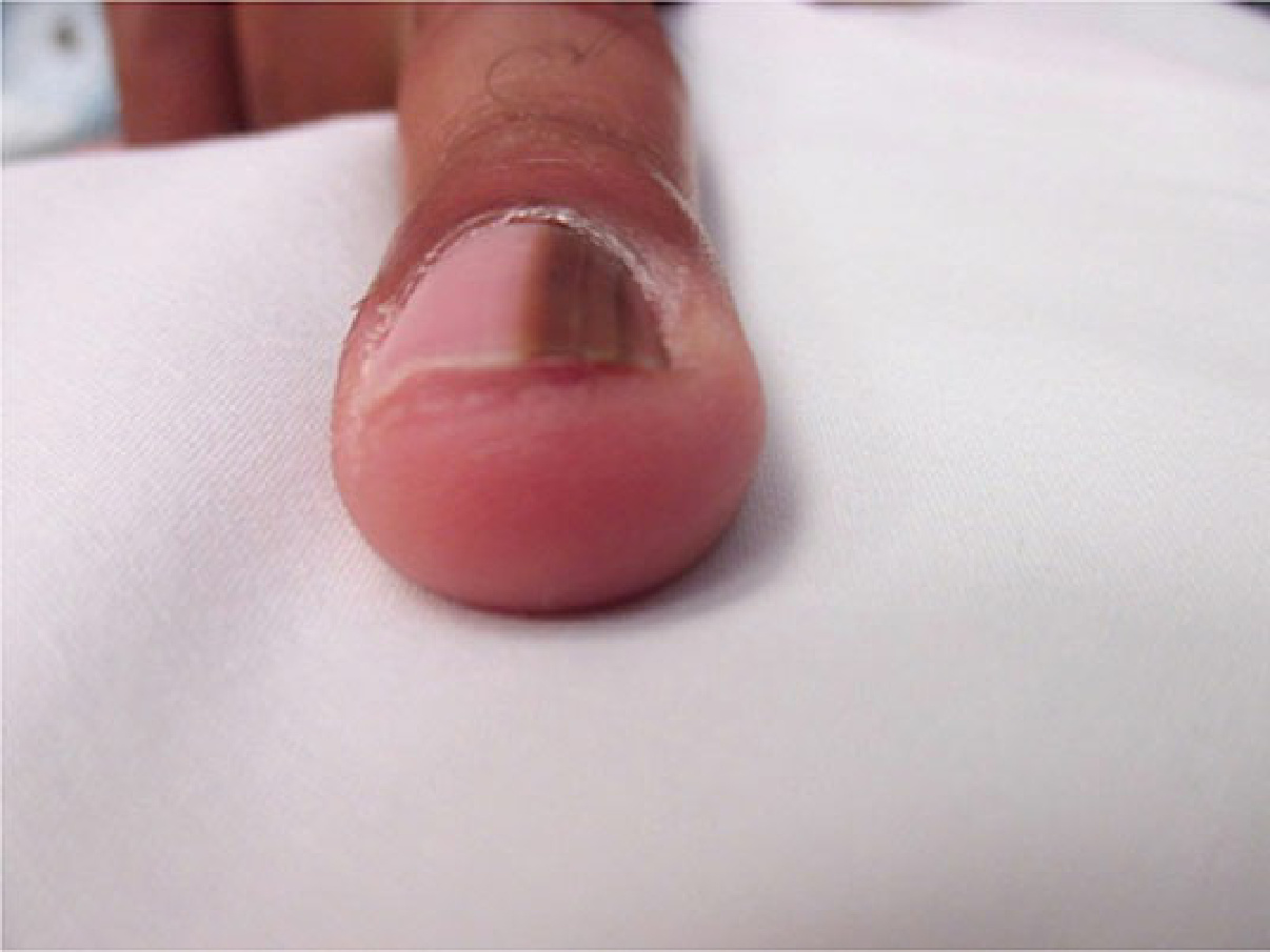
Figure 34-4. Examination of the free margin. Pigment can be seen on the surface of the nail plate which indicates that the lesion of interest is located in the proximal matrix.
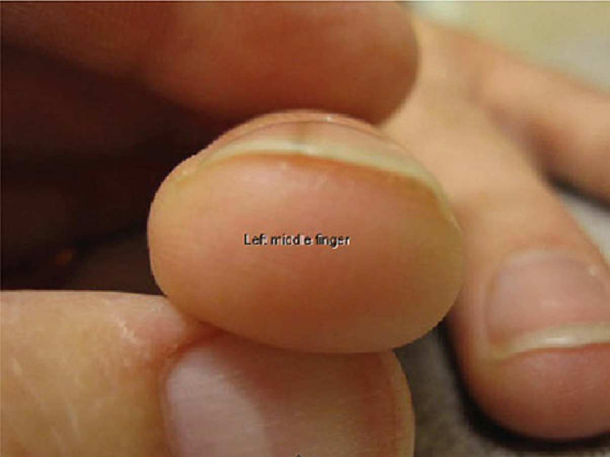
Figure 34-5. Examination of the free margin. Pigment can be seen on the undersurface of the nail plate which