Cytokeratins
Cytokeratins
Cytokeratins, or simply keratins, are intermediate filaments that play a role in the structural integrity and resilience of the epidermis. They are found in nearly all
epithelial cells, but not in nonepithelial cells such as fibroblasts, muscle cells, or neurons,18 and are therefore useful in identifying tumor cells derived from the epidermis.10 Cytokeratins are classified by their molecular weight or pH. Low– molecular-weight cytokeratins include CK7, 8, 18, 19, and 20, while CK1, 2, 5, 9, 10, and 14–17 are high–molecular-weight cytokeratins. Type I (acidic) cytokeratins include CK9–19 and type II (basic) group is composed of CK1–8. Glandular epithelium is composed of low–to intermediate–molecular-weight keratins, and squamous epithelium of primarily high–molecular-weight keratins.13
AE1/AE3 is a pankeratin stain that recognizes cytokeratins between 40 and 67kDa in molecular weight. The AE1 antibody binds to CK10, 14–16, and 19, while AE3 binds to CK1–8.18 SCC expresses CK5, 6, 8, 14, 17, and 18, while BCC expresses CK5, 14, 15, and 17.13 The AE1/AE3 cocktail will thus stain most SCCs and BCCs and can be useful in cases where there is residual tumor in areas of heavy inflammation (Fig. 30-2), perineural spread (Fig. 30-3), or extension into fascia.19,20 AE1/AE3 is also an effective choice for staining microcytic adnexal carcinoma (MAC) and sebaceous carcinoma.14
MNF116 is a monoclonal antibody that detects both low–and high–molecular-weight cytokeratins (CK5, 6, 8, 17, and 19). It shows cytoplasmic staining and is positive in all cutaneous lesions with epithelial differentiation including SCC, BCC, adnexal tumors, and Merkel cell carcinoma (MCC).21 MNF116, particularly in combination with p63, may be a sensitive stain for poorly differentiated SCC.22
CK7 is a low–molecular-weight cytokeratin and is a specific, intense stain for Paget cells as well as the immunostain of choice for EMPD.23 Sebaceous carcinoma may also express CK7.24
Cam 5.2 stains for the low–molecular-weight cytokeratin CK8, and to a lesser extent, CK7.25 It can be very useful in identifying sebaceous carcinoma and distinguishing it from benign sebaceous neoplasms, SCC, and BCC.26 Cam 5.2 staining is also positive in primary cutaneous mucinous carcinomas.14
CK20 is a low–molecular-weight cytokeratin and is frequently used in the diagnosis of MCC.
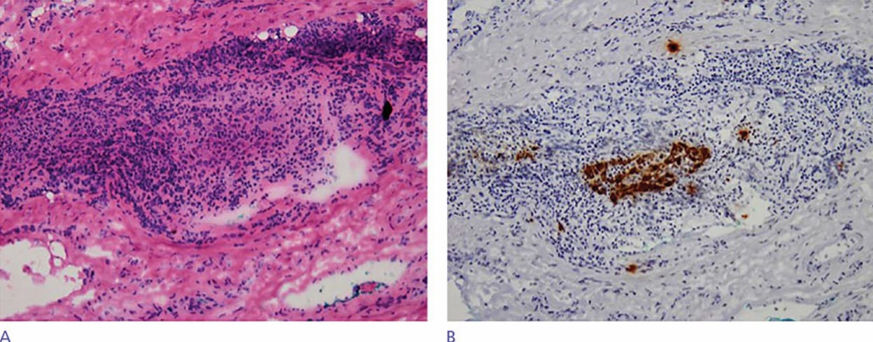
Figure 30-2. Squamous cell carcinoma with dense inflammation (400×). (A) Hematoxylin and eosin. (B) AE1/AE3 stain demonstrates carcinoma cells within inflammation. (Used with permission from Thuzar M. Shin, MD, PhD).
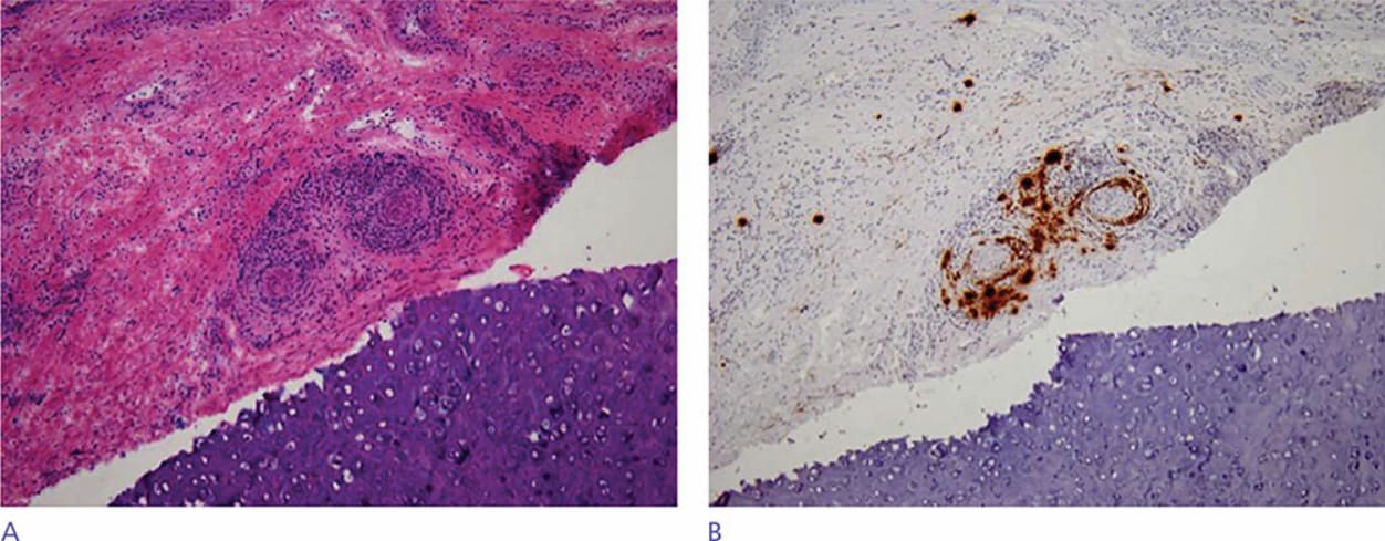
Figure 30-3. Squamous cell carcinoma with perineural invasion (100×). (A) Hematoxylin and eosin. (B) AE1/AE3 stain makes perineural tumor cells much more easily identifiable. (Used with permission from Thuzar M. Shin, MD, PhD).