Staining
Staining
Slides must be stained so that the otherwise colorless cellular material can be visualized through a light microscope. Mohs slides are generally stained with hematoxylin and eosin (H&E), toludine blue, or occasionally with special immunohistochemical stains for routine histologic analysis. H&E is the most commonly used tissue stain in MMS. This involves placing the tissue in a series of solutions: alcohol, water, xylene, hematoxylin, and eosin. In high-volume laboratories using H&E, they are placed in an automatic slide stainer to decrease the processing time (Fig. 29- 17). Toluidine blue highlights islands of BCC by metachromatically staining its surrounding mucopolysaccharides a vibrant pink.33 After the slides have been stained, they are cover-slipped. A glue-like medium is applied, followed by a thin glass cover slip that protects the tissue from being accidentally scratched off and preserves the slide for years.
If slides are moved too quickly from the fixative (usually alcohol), they will not dehydrate appropriately, resulting in suboptimal staining.
Immunohistochemistry is also used by some surgeons for treating more challenging tumors. These include cytokeratins for SCC and Paget’s disease and CD34 for
dermatofibroma sarcoma protuberans.34,35 For melanoma, frozen section interpretation can be facilitated by HMB-45,36 MART-1, and S-100 for desmoplastic subtypes,35
MITF for its superior nuclear staining,37 and mel 5.37 Overall, MART-1 is the most useful stain for melanoma.38 The cost of reagents, unavailability of automated staining equipment, additional processing time, and additional technician skill and time, have slowed the widespread adaptation of immunostains by Mohs surgeons.39 Regardless of whether immunostains are used during Mohs surgery, permanent sections may be obtained for additional confirmation of clear margins in more challenging cases. A full discussion of immunohistochemical stains for MMS may be found in Chapter 30.
During staining, a floater can be introduced from contamination of the staining solution. This floater will randomly appear (not necessarily contiguous with the specimen). Changing the staining solution regularly minimizes the introduction of floaters.
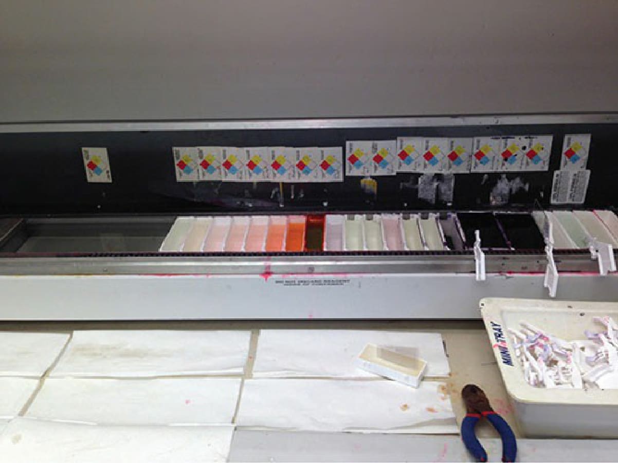
Figure 29-17. Automated hematoxylin and eosin staining device with three slides being actively stained. Once the tissue section is picked up off the roller plate by pressing one of the relabeled slides to it, the slide is stained. The stain sequence is shown above. The slide transitions from fixatives, to hematoxylin, to bluing agent, to eosin, to alcohols, to xylene. Finally, the slides are coverslipped, matched with the requisition, and presented to the Mohs surgeon for interpretation.