Cutting and slide fixation
Cutting and slide fixation
Once the specimen is embedded and frozen in the medium and mounted on the stage, it is sectioned in the microtome and placed on the slide (Fig. 29-16). The clamp usually has a ball-and-socket joint that allows for minor adjustments in specimen alignment with respect to the blade, and one must ensure proper angle of blade prior to trimming into the block. When first beginning to rotate the microtome blade, the outer layers of OCT must be cut away until reaching the actual specimen; this procedure is often termed facing the block. Upon reaching the specimen, it may require realignment to create uniform slides. If the undersurface of the specimen on the chuck is not parallel to the blade early in the cutting, incomplete sections and consequent deeper sectioning requirements may result. Once the embedding medium has been adequately trimmed, horizontal 5- to 10-micron sections (section depth varies with personal preference) are affixed to the slides. Thick sections are difficult to read as cellular detail may be obscured, although fatty tissue may call for thicker sections, while thin sections may introduce holes or tears that are visible. The first slide is of utmost importance as it represents the true margin, since the tissue is cut from the deep margin upwards toward the surface of the skin with each turn of the blade. Ensure that the technician counts the number of turns of the wheel and knows the number of microns per turn in order to properly calculate the depth of the tissue. On average, two slides are made with two to three sections per slide. Most surgeons like to see three to six sections total.32
Of note, fat does not become solid enough to cut well at temperatures that are best for cutting most other tissues. The technician may, therefore, spray the embedded fatcontaining specimen with a cryogen to lower its temperature before cutting, rather than resetting the cryostat temperature.
Nearly all problems with wrinkling and folding arise when the specimen is advanced across the blade and when picking up the wafer for placement onto the slide; therefore, the technician must ensure that the cut wafer is properly retrieved. There is a tendency for frozen tissue to curl before it is placed on a glass slide. The curling can be mitigated by the use of antiroll bars (which prevent specimens from curling out of the plane of section) or plates, and/or camel hair brushes, which are kept in the cryochamber on the brush shelf.
Dull blades can cause tearing or curling of the tissue, or even lead to chatter lines in the tissue as they cut. A microscopic nick in the edge of the blade can create a large tear in tissue. Therefore, it is important to check the blade for nicks, and to sharpen the permanent blades and move disposable blades along the blade holder throughout the day.
If the blade is not clean, floaters may be introduced by the blade. These floaters may be present on multiple slides and can therefore be difficult to recognize as floaters.
When sliding sections from the blade onto the slide, additional errors may occur. After placing each section on the slide, extra embedding medium should be wiped off the glass slide before placing the subsequent section. If the next section is placed over the embedding medium of the previous section, it may not adhere properly during fixation and can be washed off during staining. If the next section overlays embedding medium over the previous section, it may interfere with proper staining. To prevent this, when applying more than one section to a slide, it is common to begin in the upper corner and place sections diagonally across the slide.
Excessive facing of block produces false-negative and false-positive reads. If the specimen is not properly flattened, and a high area of tissue is cut away, then the true margin is forever lost. If the true margin contained tumor, a false negative may result. If the true margin was free of tumor, a false positive may result. Always use high-quality glass slides, as if glass slides are flawed tissue may not adhere properly. Charged slides (so-called plus slides) may be used to improve tissue adherence.
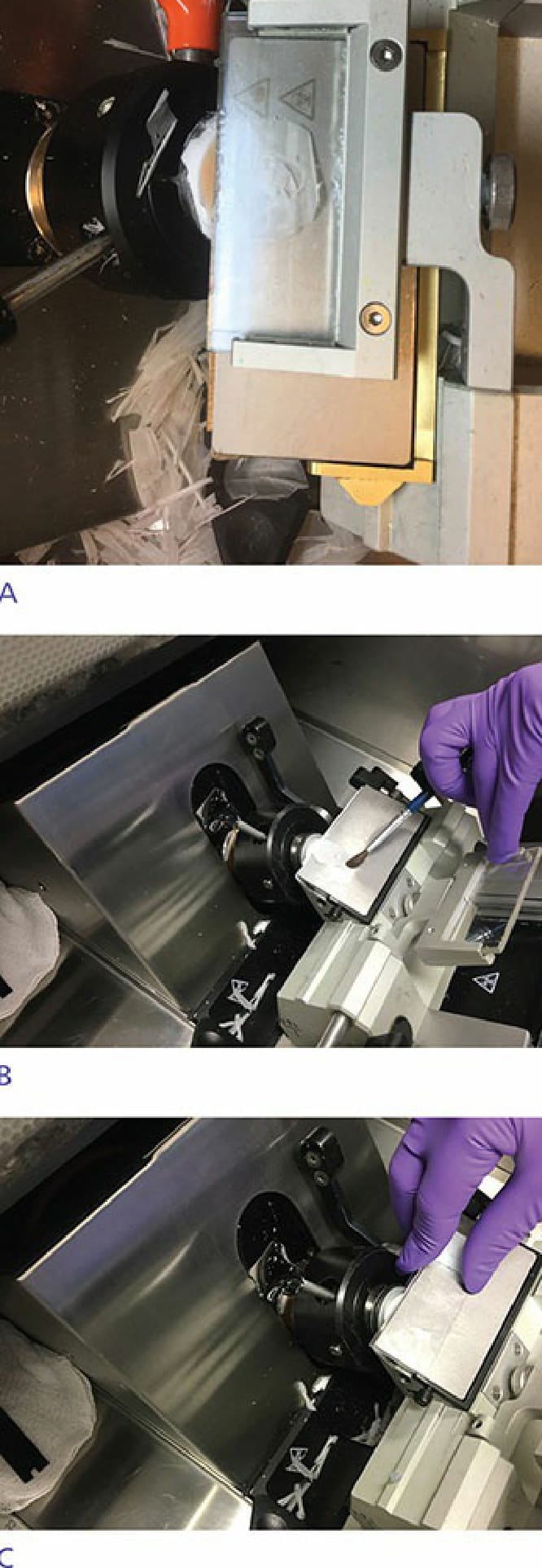
Figure 29-16. (A) Sections are taken on the roller plate by turning the cryostat wheel. (B) The brush is sitting on the roller plate. Horizontal sections are taken in a bottom-up approach so that the first few sections are a representation of the true surgical margin comprising both skin edge and deeper tissue. (C) The tissue is then transferred to a glass slide.