Sectioning and inking
Sectioning and inking
Some tumors may be sectioned into two or more pieces prior to processing; however, analysis of a single specimen with four evenly placed marks of different colors provides clear direction when correlating microscopic findings with their location on the patient. The single-section method was described by Randle et al.30 and minimizes the time needed for slide preparation and slide interpretation while also reducing the opportunity for error.31
The main factors determining when specimen subdivision is necessary are the size of microscope slides and the freezing and embedding technologies available to the technician. Convex and concave specimens, thick specimens, and specimens containing cartilage or bone usually required sectioning to be flattened into a single plane. Once appropriate sectioning has been decided, the hash marks are exaggerated, and the tissue is blotted to remove excess water (Fig. 29-9). Dyes are applied to the nicks on the excised tissue and documented on the map that was drawn at the time of excision (Fig. 29-10). Some Mohs surgeons perform the inking themselves, while other Mohs surgeon delegate this task to their technician. Analysis of the specimen with nicks and colors provides clear direction when correlating microscopic findings with their location on the patient’s defect. Inking is most helpful when the ink is placed along a significant portion of the surgical margin rather than just as an orienting dot at the specimen pole. In this situation, missing ink indicates that a margin is not fully evaluable. Most Mohs surgeons use red, blue, black, and possibly one or two other colors for this purpose.
They are represented on the Mohs map with symbols for orientation purposes.
approach the edge from the side being marked to prevent ink contamination and bleeding to unintended edges. (C) Specimen orientation, shape, reference mark location, and inking pattern are depicted on the map. In this example, XX is yellow, = is red, ….. is green, and the four vertical lines represent blue. These symbols are used so that black and white copies can still be interpreted.
Cutting tissue into more than one section introduces a higher change of mislabeling specimens or for the Mohs surgeon to incorrectly map positive margins on the incorrect section. More sections also increase the risk of false-positive and false-negative margins.31
If too much ink is used, ink may inadvertently run over and confuse the margins. This may also occur if the specimen is too wet, thus emphasizing the need to blot the tissue prior to marking (Fig. 29-11). Poor-quality ink may be difficult to read and could be washed off during tissue preparation.
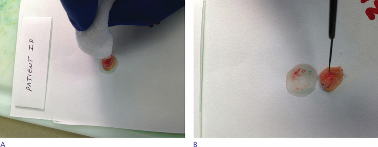
Figure 29-9. (A) The tissue is blotted to remove excess water. (B) The tissue was scored with hash marks by the Mohs surgeon during tissue removal. These small marks are enhanced by the technician to facilitate flattening the surgical margins into one plane. Along with reinforcing the existing marks, sometimes the tissue will dictate more relaxing cuts to be able to lay the entire specimen flat. When this cannot be accomplished by this technique, the tissue will be divided into sections to assure that the tissue sits on one plane, albeit in pieces.
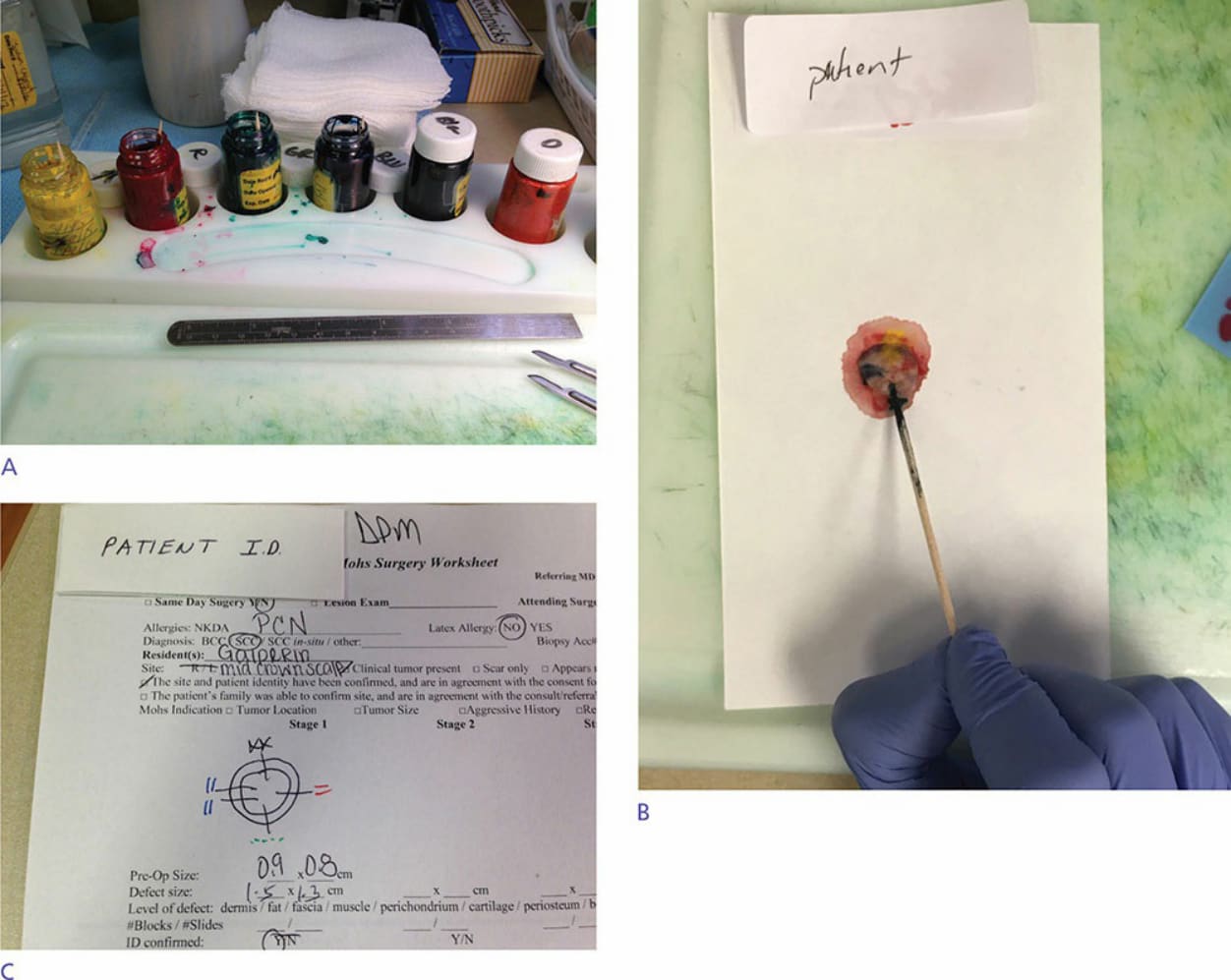
Figure 29-10. (A) Commercially available dye system with applicators are placed in a stable holder. Once the tissue has been satisfactorily relaxed, ink is applied to the hash marks on the tissue to assist the Mohs surgeon in orientation if more stages are needed. (B) A cotton-tipped applicator is used to mark the tissue with tissue dye, which remains on the tissue through histochemical staining. When applying different colored inks to nearby tissue edges, it is good to
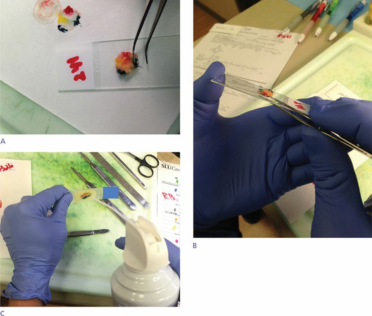
Figure 29-11. (A,B) The inked, relaxed tissue is placed on a labeled slide, deep margin down. Using forceps, the edges of the tissue are teased down flat on the slide. (C) Many times freeze spray is needed to help hold the tissue to the slide in a flat configuration. Flattening is important so that the epidermis, dermis, and subcutaneous fat lie in the same plane.