Technique
Technique
The donor site is first scrubbed with antiseptic and anesthetized. The surgical defect is then measured in both horizontal and vertical dimensions and a graft template is drawn with a surgical marking pen on the donor site. If the graft donor site is to be closed primarily, the standing cones to be removed can be drawn at the same time. It is recommended to oversize the harvested graft 10% to 20% to account for tissue contraction and ensure a final graft that is sufficiently sized for the surgical defect (Fig. 28-4).
layered closure.
A no. 15 Bard-Parker scalpel is used to excise the donor graft tissue with or without the planned standing cones. If the surgical defect to be repaired is a defect from Mohs micrographic surgery, beveled edges are usually present in the wound bed. If the beveled edges are maintained, the graft should be harvested at a 45-degree angle for optimal fit.25 If the graft is excised at the standard 90-degree angle, the wound edges of the Mohs surgical defect should be trimmed to reflect right angles, so the graft may optimally fit in place.
The donor site is excised down to the level of the adipose tissue, with the exception of the conchal bowl graft, which is excised to the level of full dermal thickness down to cartilage, leaving perichondrium intact. Immediately after excision, the graft is placed in a small sterile basin containing sterile saline, and hemostasis is achieved at the graft donor site.
The graft is then placed epidermis down with subcutaneous fat exposed over the gloved nondominant hand or over a gauze pad in the nondominant hand. The harvested graft is traditionally de-fatted with curved iris scissors until white shiny dermis is exposed over the entire skin graft. It should be noted that although de-fatting is a standard practice in dermatologic surgery to minimize the risk of graft necrosis, retaining 1 to 5 mm of subcutaneous fat on FTSGs may be considered with pleasing aesthetic results, particularly for the repair of deeper defects on the lower one-third of the nose.26
The skin edges of the recipient wound bed are then undermined to minimize potential postoperative pincushion deformity. The graft is accurately positioned over the wound bed, and simple interrupted sutures are placed at opposite ends of the graft to secure it in place. Meticulous hemostasis must be performed in the wound bed prior to graft placement to ensure that hematoma does not interfere with the graft’s contact with the nutrient-rich wound bed. Depending on graft location, 6-0 polypropylene, 5-0 polypropylene, or 5-0 fast-absorbing gut sutures are favorable sutures to secure the graft.
Iris scissors are used to trim the graft to the exact size of the wound bed, and additional simple interrupted sutures are placed to secure the graft. The suture is first placed through the graft and then through the donor skin to maximize accurate approximation and minimize trauma to and displacement of the graft. Sutures should be placed down to the level of the reticular dermis of the graft and donor site skin to anchor the graft to the wound bed for optimal graft survival and to reduce the risk of depressed scar lines.27 Central basting sutures may also be placed to maximize graft contact with the wound bed. For larger grafts, a running suture may also be used once several anchoring sutures are placed evenly around the periphery of the graft.
The donor site is then repaired in a standard fashion using a layered closure technique. If a conchal bowl graft is harvested, the graft donor site is left to heal by second intention. If a delayed FTSG is performed, the graft is placed over the granulating wound bed 2 to 4 weeks after surgery. Before graft placement, the wound bed is first curetted down to a healthy base, and the skin edges are freshened by excising 1- to 2-mm margin of the wound edge.19
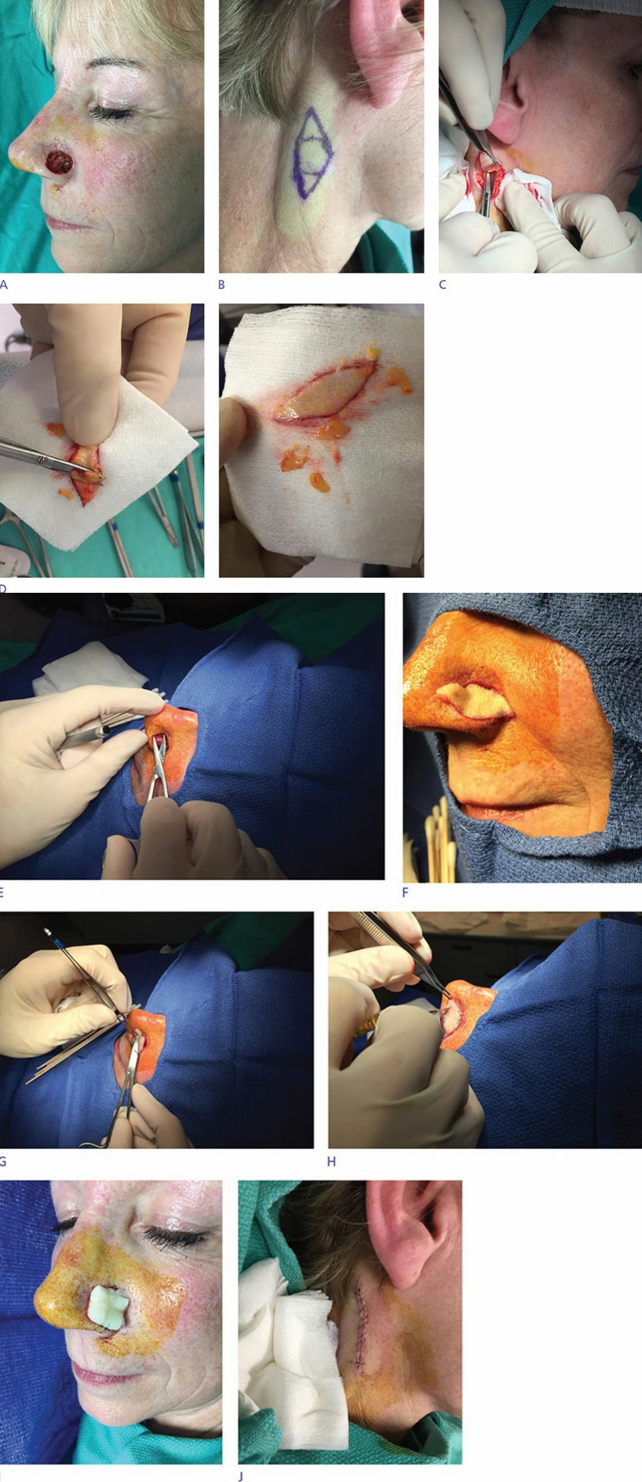
Figure 28-4. Step-by-step FTSG repair. (A) A surgical defect on the left nasal ala is present after Mohs micrographic surgery for a basal cell carcinoma. (B) The defect is measured and a template is drawn on the neck, which has been selected as the FTSG donor site. (C) The donor site skin is excised in a standard fashion. (D) Iris scissors are used to de-fat the FTSG until shiny white dermis is visible across the graft. (E) The surgical defect is undermined to minimize postoperative pincushion deformity. (F). The FTSG is placed on the recipient wound bed. (G) The graft is sutured in place with anchoring sutures. (H) The FTSG is trimmed to perfectly fit the surgical defect. (I) Remaining sutures are placed (not pictured) and a bolster dressing is placed as desired. (J) The FTSG donor site is repaired with a standard