Flap design
Flap design
When designing any flap, it is important to consider function, free margins, and form. The PFF cannot simply be placed on the distal nose without proper structural support to prevent collapse of the nasal valve. Cartilage grafts provide structural support, prevent alar retraction, and also help to restore the normal form and contour of the nose.2,3 The grafts are often harvested from the conchal bowl or antihelix for convenience, though nasal septum and costal sources can also be considered. If the nasal lining is not intact, mucosal flaps, hinge flaps, bipedicle septal flaps, or inverted full-thickness skin grafts can be used.4–7 A three-stage fold-over PFF is an excellent option for repair (Fig. 26- 1A,B).
For defects involving >50% of a cosmetic subunit, enlarging the operative wound to encompass the entire subunit may provide a superior aesthetic outcome. A template of
the defect is made using gauze, Telfa, steri-strips, or foil from a suture packet prior to excision of the remaining subunit (Fig. 26-2). The template is then rotated 180 degrees onto the forehead (Fig. 26-3). The contralateral forehead is often chosen for the flap as this rotates with less torsion. If the defect is midline, either side of the forehead can be used, and laterality is decided based on the quality of the skin and the lack of any other local suspicious lesions. The distance from the anterior hairline to the superior orbital rim is measured to determine the length of the flap (Fig. 26-4A,B). If the reach is not adequate, the flap may extend onto the scalp or below the superior orbital rim. A paramidline flap design located 1.2 cm lateral to the midline of the glabella may be beneficial when longer flaps are needed, as extending the pedicle inferiorly does not affect the brow.8 Extension onto the scalp will transfer terminal hairs to the nose, though this should not prevent the use of this repair, as creating a normal-appearing nose takes precedence. The flap can be thinned distally to remove hair follicles, or the patient can shave, epilate, or undergo laser hair removal or electrolysis postoperatively.
The vascular supply of the pedicle is based on the supratrochlear artery, which may be determined based upon anatomical landmarks or using a handheld Doppler ultrasound. No difference in flap survival or complications was seen when comparing these two techniques.8 The most prominent glabellar frown line and 6 mm of lateral skin reliably corresponds to the location of this artery, and can be used in lieu of Doppler localization.9 The width of the pedicle is rarely greater than 1.5 cm, as wider pedicles
may limit the flap’s mobility and impair blood supply due to greater torsion and compression of the artery.
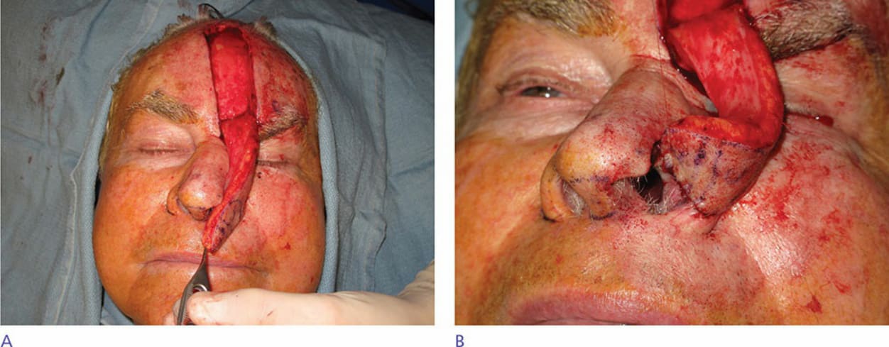
Figure 26-1. (A,B) Recreating the intranasal lining using a three-stage fold-over paramedian forehead flap.
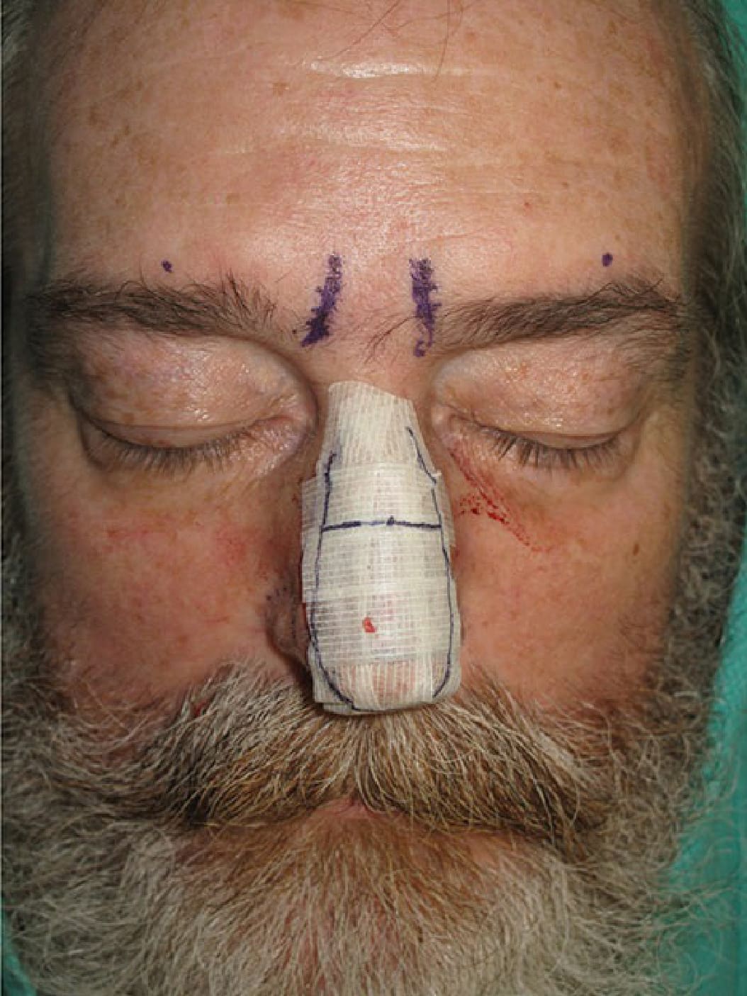
Figure 26-2. A three-dimensional template is created using steri-strips.
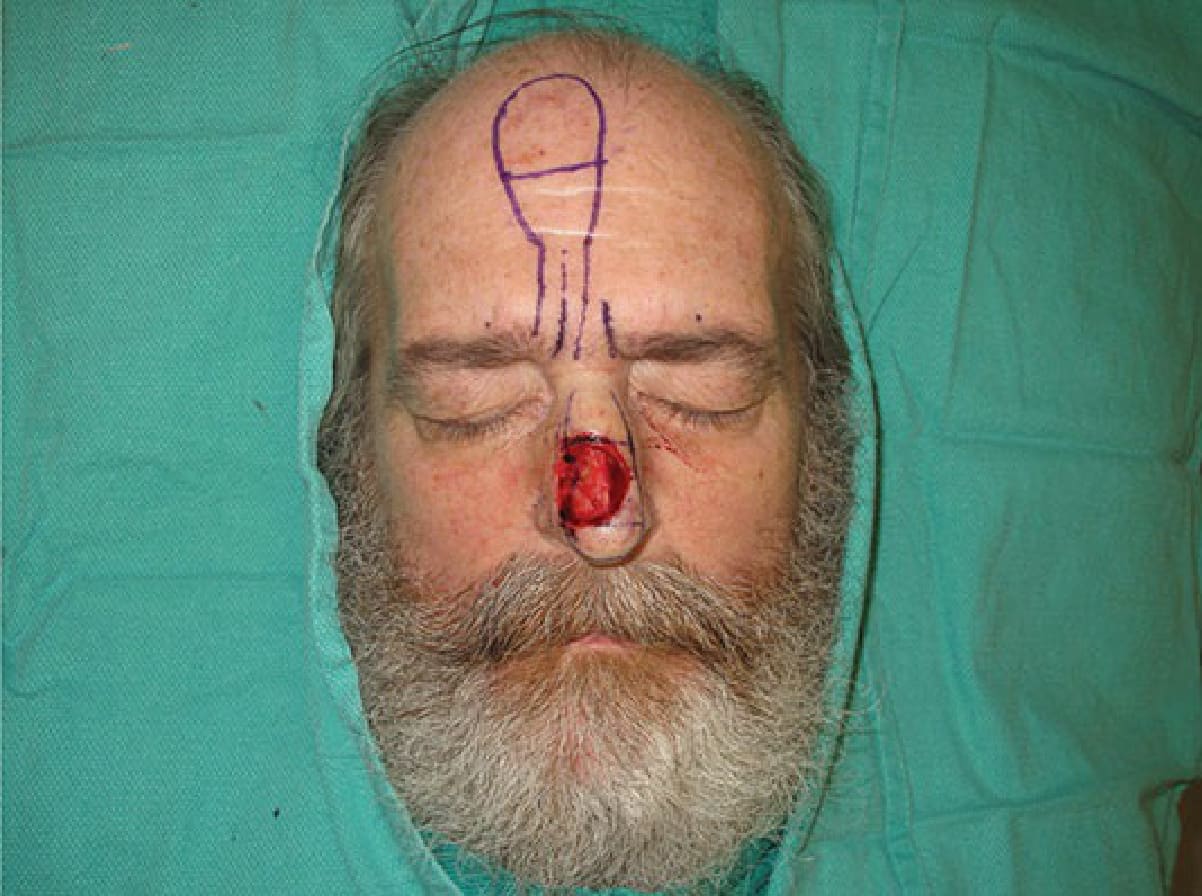
Figure 26-3. The template is inverted onto the forehead. The pedicle is marked 6 mm to each side of the most prominent corrugator crease.
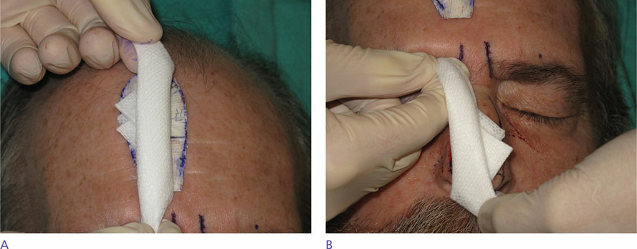
Figure 26-4 (A,B) The distance from the base of the pedicle to the top of the template is measured with gauze. The gauze is then rotated toward the nose to confirm flap reach.