Suturing technique considerations
Suturing technique considerations
Unlike certain other local flaps, such as island pedicle flaps and bilobed flaps, advancement flaps are rarely associated with postoperative pin-cushioning. Moreover, the straight limbs of an advancement flap are often easier to camouflage along cosmetic subunit junctions. The surgeon should be careful to avoid hypereverting scars that are to be camouflaged in concave cosmetic subunit junctions, such as the nasolabial fold, as hypereversion of these areas may be associated with residual ridging. The base of an advancement flap is generally under minimal to no tension, so undesirable eversion in this location may take longer to settle (Fig. 21-8). To mitigate this risk, suturing techniques may be adjusted accordingly, and simple buried sutures (that may encourage slight inversion) may be used in lieu of everting techniques such as the set-back suture
or buried vertical mattress suture. If needed, suspension sutures or inverting techniques may be used as well; for a full discussion of tailored suturing approaches, see Chapter 13.
Anchoring procedures and SMAS plication for periocular flaps
The lower eyelid is vulnerable to ectropion from vertical tension, even if the defect does not directly involve the eyelid margin.2 To avoid eyelid malposition, the lower eyelid may be anchored to the periosteum of the lateral orbital rim for support (Fig. 21- 9). Laterally based suspension sutures in periocular reconstruction advance the tissue around the eyelid defect to the lateral orbital rim.14 SMAS plication may be occasionally helpful to avoid eyelid malposition and counteract the weight of a flap or gravitational pull for periocular advancement flaps.15 The degree of SMAS lift may be as small as a lateral suborbicularis oculi fat (SOOF) elevation or as large as a modified lateral midface lift, depending on the size of the defect and the individual patient’s anatomy. When performed, these techniques are often executed in concert with a canthoplasty.
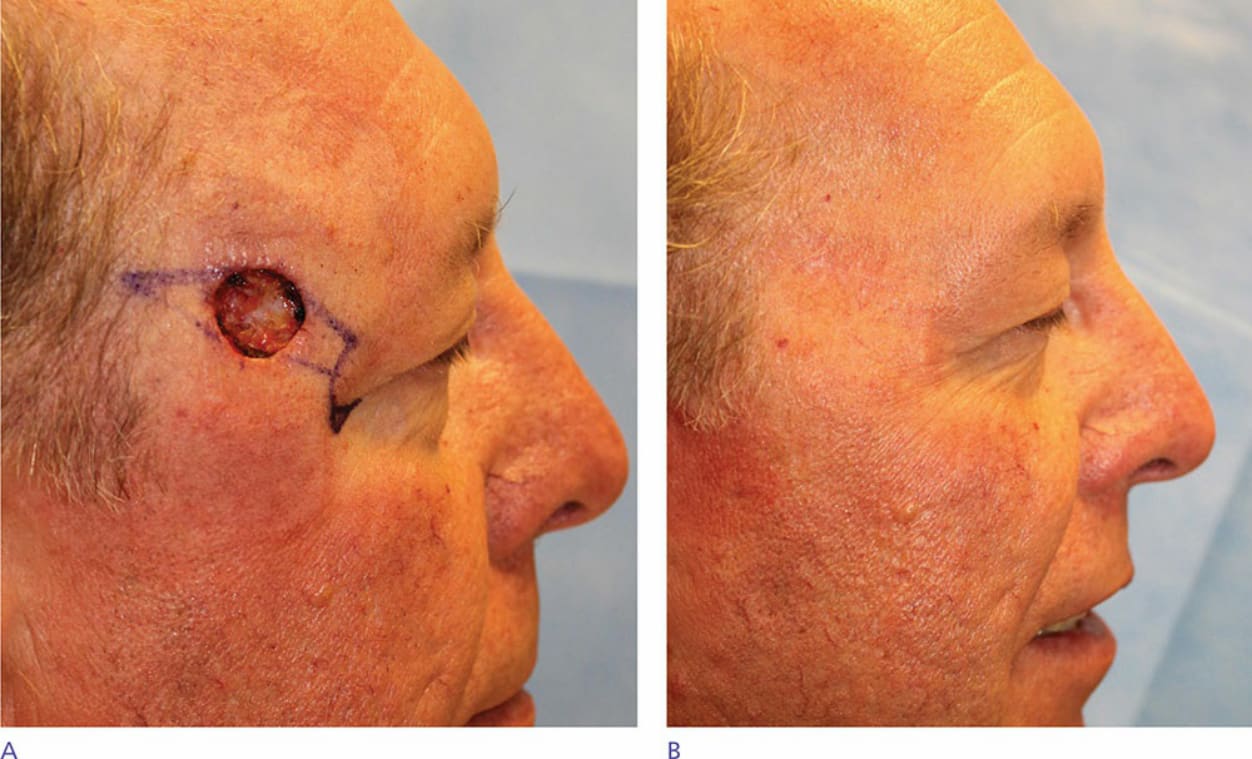
Figure 21-8. An advancement flap was used to reconstruct this defect on the lateral suprabrow (A). The incision line used to displace the standing cone is typically under minimal tension. Eversion along this line was persistent at 8 months postoperatively and can take substantial time to resolve spontaneously (B).
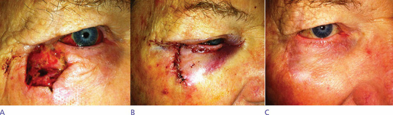
Figure 21-9. Defect photo (A) after resection of a tumor on the lateral canthus and lower eyelid. The wound was repaired with an O-to-T advancement flap (B). To prevent downward traction on the eyelid, an external canthoplasty and a SMAS lift were performed to elevate the midface and prevent eyelid retraction (B). The patient had excellent apposition between the globe and the lower eyelid postoperatively (C).