Shave
Shave
Biopsies using a shave technique are commonly used to sample neoplastic lesions, but may also be useful in the setting of inflammatory dermatoses if the key histologic features are present in the epidermis and papillary dermis. Engineered sharps (Fig. 14-
2) should be considered whenever possible to reduce the risk of injury to members of the care team, though currently available engineered sharps are less rigid that traditional double-edged razor blades and often cannot substitute. Thus most dermatologic surgeons
continue to prefer the use of either double- or single-edged razor blades. Others prefer the use of a number 15 or number 10 blade followed by curettage to smooth the wound edges.
The technique. The skin is pulled taught in all directions by the surgeon, and an assistant if needed. This allows for more precise sampling of the tissue, a smoother wound edge, and quicker sampling. A shave that won’t end, producing a tail, is usually the result of failure to pull the skin taught. The blade is initially angled slightly downward to breach the epidermis, carried horizontally to obtain the specimen, then angled upward to complete the procedure. A fairly rapid but gentle side-to-side motion allows the blade to move smoothly through the tissue without causing ridges. A tumid technique has also been described which involves ballooning of soft papules such as benign nevi and neurofibromas with a mixture of lidocaine diluted with bacteriostatic 0.9% sodium chloride to create a more rigid structure and facilitate shave removal.6
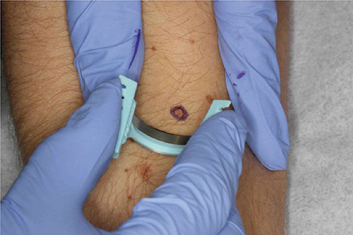
Figure 14-2. Shave biopsy technique. The skin is held taut with the nondominant hand, and the fourth finger of the dominant hand may provide additional stabilization.