Techniques
Techniques
INTRODUCTION
The biopsy of tissue for histologic examination remains the most informative and costeffective test in medical practice and one of the most common procedures performed by dermatologists. Usually, a visible lesion is biopsied, but multiple random skin biopsies from the abdomen and thighs may be helpful in the setting of angiotropic lymphoma presenting as fever of unknown origin1, to obtain tissue for fibroblast culture to assess for a genodermatosis,2 or to assess the possibility of immunobullous disease in a patient with generalized pruritus. Reflectance confocal microscopy can be useful to localize the best area to biopsy in a large, poorly defined lesion.3 A range of biopsy techniques are available to the clinician (Fig. 14-1).
All tissues removed during a surgical procedure should be submitted for histopathologic examination unless specifically exempted by an organization’s Tissue Committee policy. Such policies typically exempt certain tissues of limited diagnostic value such as femoral head, teeth, nails, and cyst contents. Skin tags are sometimes on the exempt list, but caution is advised, as infundibulocystic basal cell carcinoma,
neurofibromas, and fibrofolliculomas may have a tag-like appearance. Any unusualappearing tag should be submitted for examination, even if Tissue Committee policy exempts them.
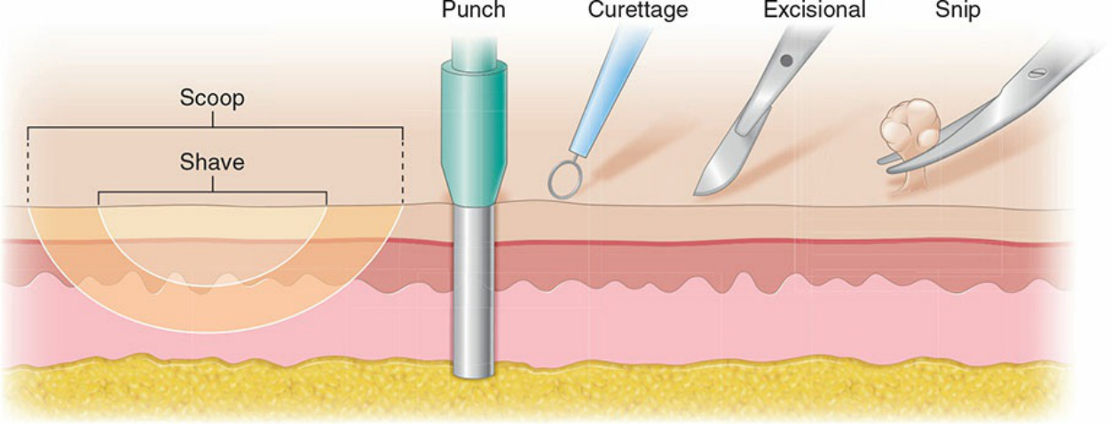
Figure 14-1. A range of biopsy techniques are available.