Temple
Temple
The superficial temporal artery travels within the layers of the superficial temporal
fascia.
Rich anastomosis occurs between branches of the superficial temporal artery and
supraorbital artery.
The auriculotemporal nerve runs deep and posterior to the superficial temporal
artery.
The frontal branch of the facial nerve is most vulnerable as it crosses the
zygomatic arch as a single trunk en route to the deep surfaces of the frontalis muscle.
The temporal fossa contains a relatively sparse amount of subcutaneous tissue devoid of muscles of facial expression, with the exception of traversing fibers of the orbicularis oculi muscle and even fewer fibers of the anterior auricular muscle. Two distinct layers of fascia are contained within this unit. The deep temporal fascia, which is a continuation of the investing fascia containing the deeper temporalis muscle as it becomes continuous with the periosteum of the skull; and the superficial temporal fascia, which is a continuation of the SMAS as it connects to the galea aponeurotica (Fig. 1-12). In this region, the superficial temporal fascia is of anatomical and subsequent surgical importance as it contains within its layers key vascular and neural structures as they traverse between the fascial layers. The superficial temporal artery along with its branches and sensory nerves, including the auriculotemporal nerve, can be accessed within the layers of the superficial temporal fascia (Fig. 1-12). The motor branches of the facial nerve remain deep to the superficial temporal fascia as they course toward the deep surface of the orbicularis oculi and frontalis muscles (Fig. 1-6). The superficial temporal fascia forms a continuous layer with the galea aponeurotica, but splits medially to enclose the frontalis and orbicularis oculi muscles and laterally the superficial periauricular fibers. Inferiorly, the superficial temporal fascia is adherent to the zygomatic arch. Immediately adjacent to the superficial layer of the superficial temporal fascia, the subcutaneous fatty layer separates it from the overlying dermis. Fibrous septa create a more taut area as one moves toward the scalp, with relatively greater laxity just above the zygomatic arch. Numerous cutaneous vessels and nerves lie in this interval between the fat and fascia, which is important to remember when undermining in this area. The deep layer of the superficial temporal fascia glides over the loose connective tissue of the deep temporal fascia, deep to which the temporalis muscle can be visualized (Fig. 1-12).
The primary source of vascular supply to the temple comes from the superficial temporal artery, a terminal branch of the external carotid artery. The superficial temporal artery emerges from the superior pole of the parotid gland as it pierces the parotid fascia anterior to the tragus (Fig. 1-12). Inferior to it, the transverse facial artery runs below and in line with the zygomatic arch. The artery is accompanied by the corresponding veins, and usually divides anteriorly into anterior and posterior branches, with two or sometimes three significant sized pedicles. The anterior branch follows a distinct tortuous course, especially prominent in elderly patients, to supply the temple and the temporal scalp region. Branches anastomose freely with the posterior parietal branches as well as contributions from the supraorbital artery. From an anatomical standpoint, it is important to note that while the superficial temporal artery lies within the layers of the superficial temporal fascia, the corresponding veins are located within the subcutaneous layer. As the arteries continue toward the scalp, they also come to lie within the subcutaneous plane just above the superficial temporal fascia.
Sensory innervation to the temple is achieved via the maxillary and mandibular divisions of the trigeminal nerve. The auriculotemporal nerve (Fig. 1-13) travels posterior and deep to the superficial temporal artery and branches as it runs within the same fascial plane as the artery as they proceed toward the scalp. The skin adjacent to the lateral canthus is supplied by a branch of the maxillary artery, with the zygomaticotemporal nerve emerging from the lateral orbital wall. Additionally, the zygomaticotemporal nerve innervates an area of scalp between the territories of the auriculotemporal and supraorbital nerves (Fig. 1-8). Emerging from the superior pole of the parotid gland, the temporal branch of the facial nerve crosses superficial to the
zygomatic arch as a single branch within the superficial temporal fascia increasing its vulnerability to surgical injury (Fig. 1-14). With the use of surface anatomical landmarks, the temporal branch may be visualized along a line 0.5 cm below the tragus to a point approximately 1.5 cm superior to the lateral edge of the eyebrow. The temporal branch of the facial nerve supplies the frontalis muscle from the deep lateral edge with few branches contributing to fibers of orbicularis oculi and those of surrounding muscles of facial expression.
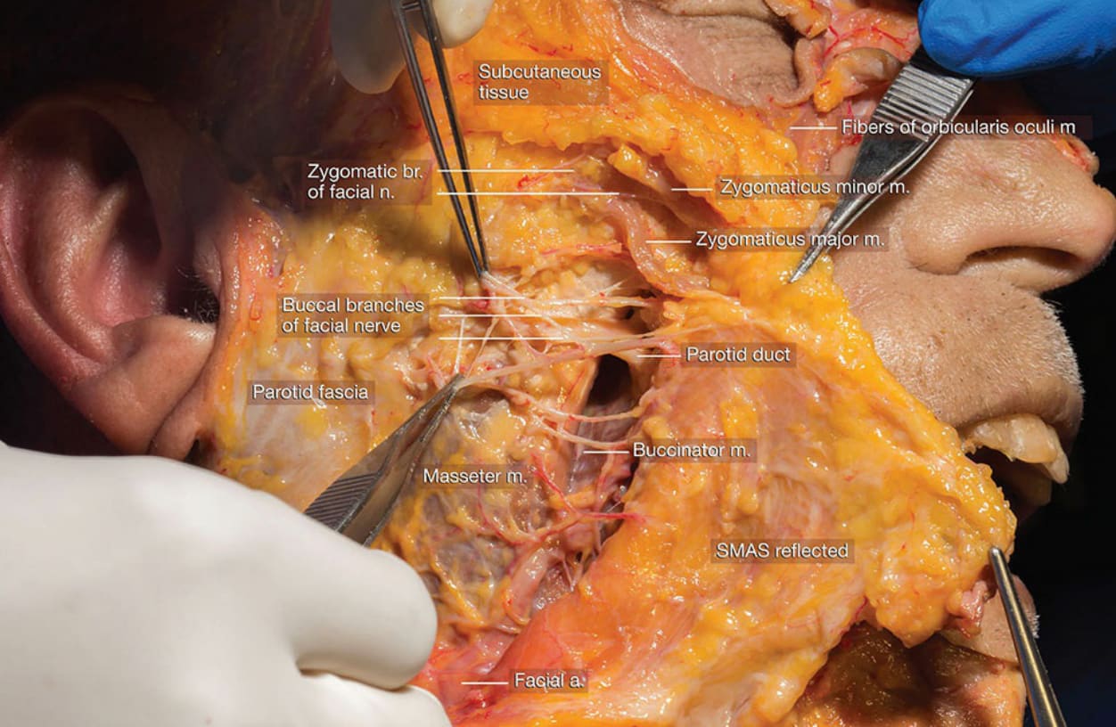
Figure 1-6. Dissection of facial nerve deep to SMAS.
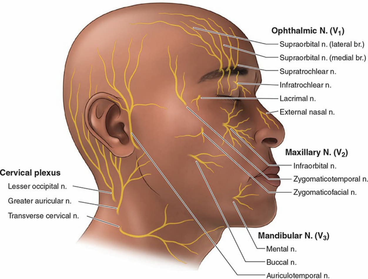
Figure 1-8. Diagram illustrating distribution patterns of sensory branches of the trigeminal nerve.
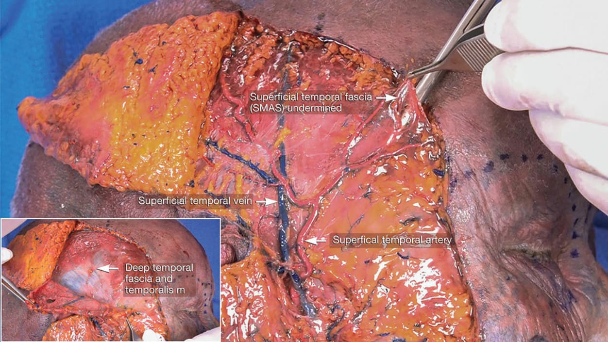
Figure 1-12. Dissection of superficial temporal fascia reflected to show superficial temporal vessels.
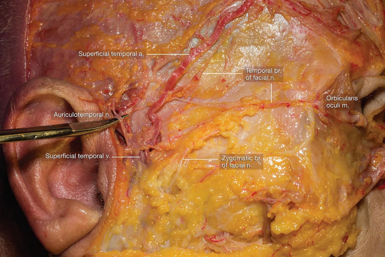
Figure 1-13. Dissection of temporal region highlighting the auriculotemporal nerve.
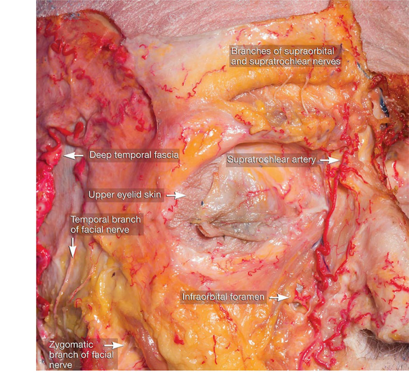
Figure 1-14. Dissection demonstrating relationship between superficial temporal artery and temporal branch of the facial nerve.