Incisional biopsy
Incisional biopsy
Incisional biopsies allow the surgeon to have full control over the size, shape, and depth of the specimen and are typically followed by straight-line closure. Panniculitis is best diagnosed by an incisional biopsy, although double- punch and other related techniques have also been used.8 Large melanocytic lesions are also often approached in this manner.
The technique. A straight line or elliptical incision is made and the desired portion of tissue is removed (Fig. 14-11). The resulting defect is then similar to any other created in excisional surgery (Fig. 14-12), and the wound is closed with simple or layered closure.
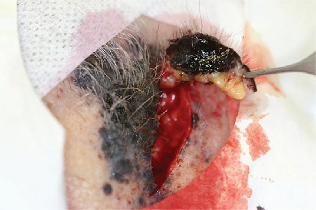
Figure 14-11. An incisional biopsy is useful particularly when a deep melanoma is being sampled to assess the depth of invasion.
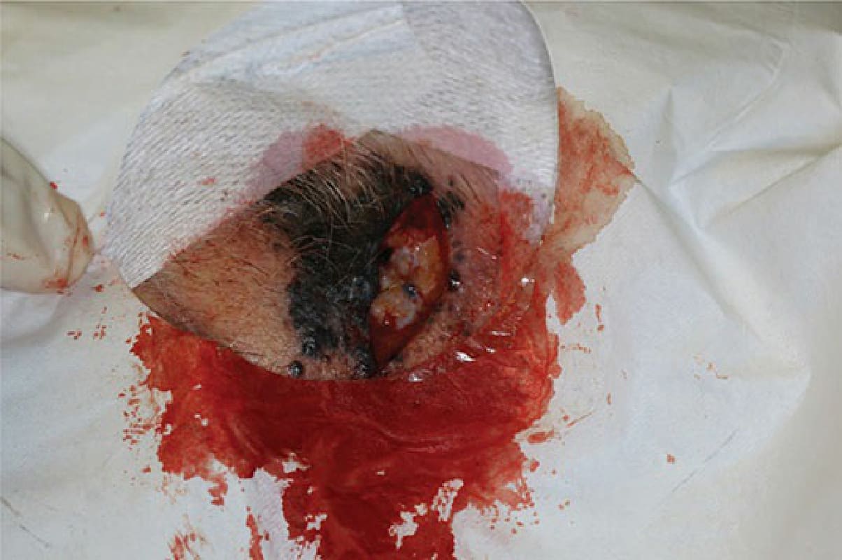
Figure 14-12. The resultant defect may then be approached in a similar fashion to any other excisional surgery.