Saucerization
Saucerization
When performed properly, saucerization, or scoop shave removal, results in an eponymous saucer-shaped specimen with complete extirpation of the lesion (Fig. 14-3). Saucerization may remain in the dermis or be scooped to subcutaneous tissue.
The technique. The overall technique is very similar to a shave biopsy, but the blade is angled more steeply toward the center of the biopsy site, then advanced with a similar rapid but gentle side-to-side motion and a forward scooping motion. Some clinicians prefer to score the skin around the target lesion top aid with an aggressive saucerization. Often, pinpoint-bleeding dermis is visible immediately after a saucerization biopsy (Fig. 14-4).
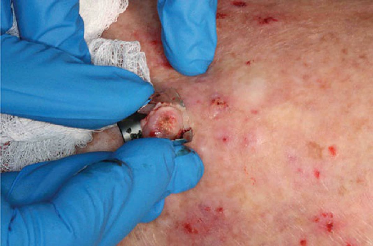
Figure 14-3. Saucerization biopsy is essentially a deep shave biopsy. This is a preferred technique for suspicious pigmented lesions.
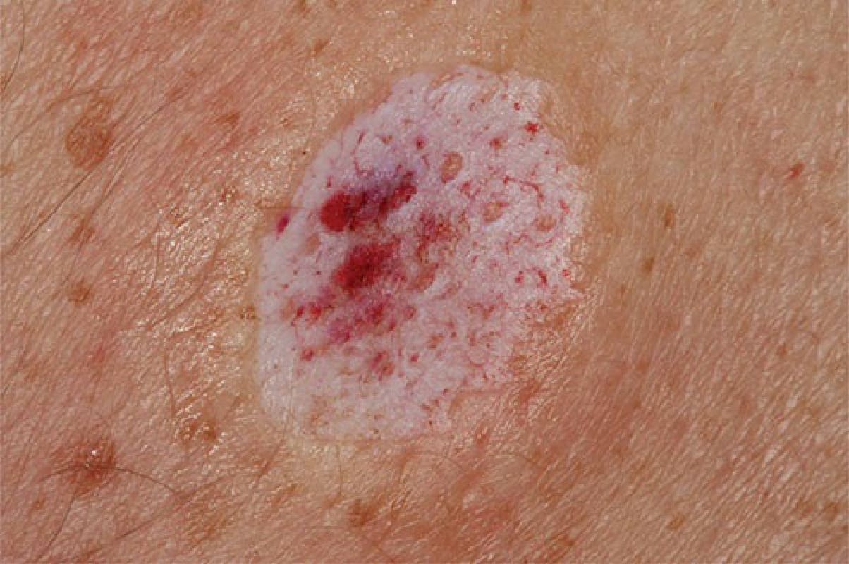
Figure 14-4. Often, pinpoint-bleeding dermis is visible immediately after a saucerization biopsy.